Department of Ophthalmology, Saarland University Medical Center, Kirrberger Str. 100, D-66424, Homburg, Saar, Germany.
Dr. Rolf M. Schwiete Center for Limbal Stem Cell and Aniridia Research, Saarland University, Homburg, Saar, Germany.
Graefes Arch Clin Exp Ophthalmol. 2021 May;259(5):1225-1234. doi: 10.1007/s00417-020-05058-z. Epub 2021 Jan 14.
To analyze the effect of riboflavin UV-A illumination on mRNA and protein expression of healthy (HCFs) and keratoconus human corneal fibroblasts (KC-HCFs), concerning the inflammatory markers NF-κB, iNOS, IL-6, and collagen 1 and 5 (Col 1/Col 5).
Keratocytes were isolated from healthy (n = 3) and keratoconus (KC) corneas (n = 3) and were cultivated in basal medium with 5% fetal calf serum, which resulted in their transformation into human corneal fibroblasts (HCFs/KC-HCFs). Cells underwent 0.1% riboflavin UV-A illumination for 250 s (CXL). NF-κB, iNOS, IL-6, Col 1, and Col 5 expression was investigated by qPCR and Western blot analysis. IL-6 concentration of the cell culture supernatant and cell lysate was determined by ELISA.
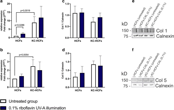
In untreated KC-HCFs, NF-κB (p = 0.0002), iNOS (p = 0.0019), Col 1 (p = 0.0286), and Col 5 (p = 0.0054) mRNA expression was higher and IL-6 expression was lower (p = 0.0057), than in healthy controls. In HCFs, CXL led to an increased NF-κB (p = 0.0286) and IL-6 (p = 0.0057) mRNA expression. The IL-6 concentration in the cell culture supernatant was increased in HCFs (p = 0.0485) and KC-HCFs (p = 0.0485) after CXL. CXL increased intracellular IL-6 concentration only in KC-HCFs (p = 0.0357). In the HCF group (p = 0.0286), an increased Col 1 mRNA expression after CXL could be observed.
Our study confirmed altered gene expression in untreated KC-HCFs compared to untreated HCFs. Riboflavin UV-A illumination affected gene expression only in HCFs. Increased IL-6 concentration in the cell culture supernatant and cell lysate indicate a secondary inflammatory response of HCFs and KC-HCFs to riboflavin UV-A illumination.
分析核黄素紫外线 A 照射对健康(HCFs)和圆锥角膜人角膜成纤维细胞(KC-HCFs)的 mRNA 和蛋白质表达的影响,研究炎症标志物 NF-κB、iNOS、IL-6 和胶原 1 和 5(Col 1/Col 5)。
从健康(n=3)和圆锥角膜(KC)角膜(n=3)中分离角膜细胞并在含有 5%胎牛血清的基础培养基中培养,使其转化为人角膜成纤维细胞(HCFs/KC-HCFs)。细胞接受 0.1%核黄素紫外线 A 照射 250 秒(CXL)。通过 qPCR 和 Western blot 分析研究 NF-κB、iNOS、IL-6、Col 1 和 Col 5 的表达。通过 ELISA 测定细胞培养上清液和细胞裂解液中的 IL-6 浓度。
在未经处理的 KC-HCFs 中,NF-κB(p=0.0002)、iNOS(p=0.0019)、Col 1(p=0.0286)和 Col 5(p=0.0054)mRNA 表达更高,IL-6 表达更低(p=0.0057),与健康对照组相比。在 HCFs 中,CXL 导致 NF-κB(p=0.0286)和 IL-6(p=0.0057)mRNA 表达增加。HCFs 和 KC-HCFs 经 CXL 后,细胞培养上清液中的 IL-6 浓度增加(p=0.0485)。CXL 仅增加 KC-HCFs 细胞内的 IL-6 浓度(p=0.0357)。在 HCF 组(p=0.0286)中,CXL 后观察到 Col 1 mRNA 表达增加。
本研究证实了未经处理的 KC-HCFs 与未经处理的 HCFs 相比,基因表达发生了改变。核黄素紫外线 A 照射仅影响 HCFs 的基因表达。细胞培养上清液和细胞裂解液中 IL-6 浓度的增加表明 HCFs 和 KC-HCFs 对核黄素紫外线 A 照射的二次炎症反应。