Department of Cell Biology, University of Oklahoma Health science Center, Oklahoma City, Oklahoma, USA.
Department of Ophthalmology, Aarhus University Hospital, Aarhus C, Denmark.
Sci Rep. 2017 Oct 2;7(1):12517. doi: 10.1038/s41598-017-12598-8.
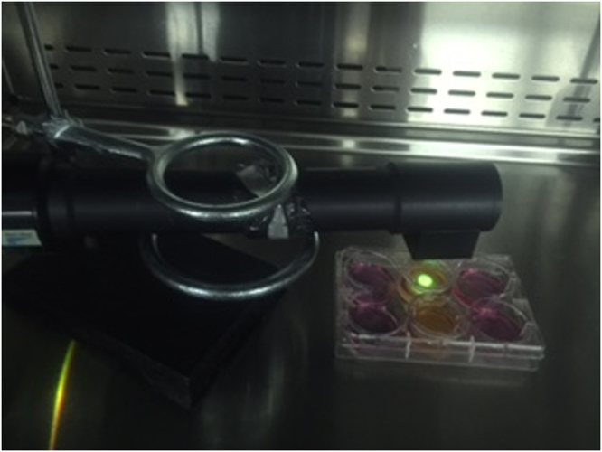
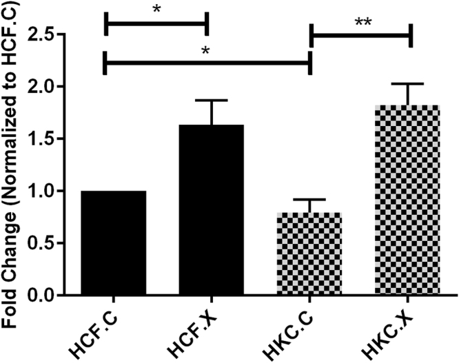
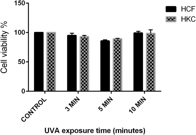
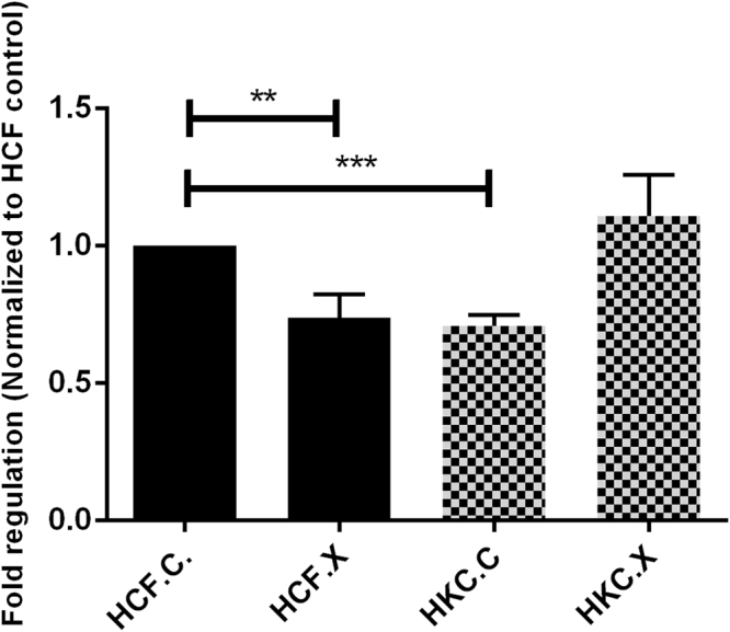
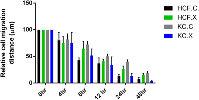
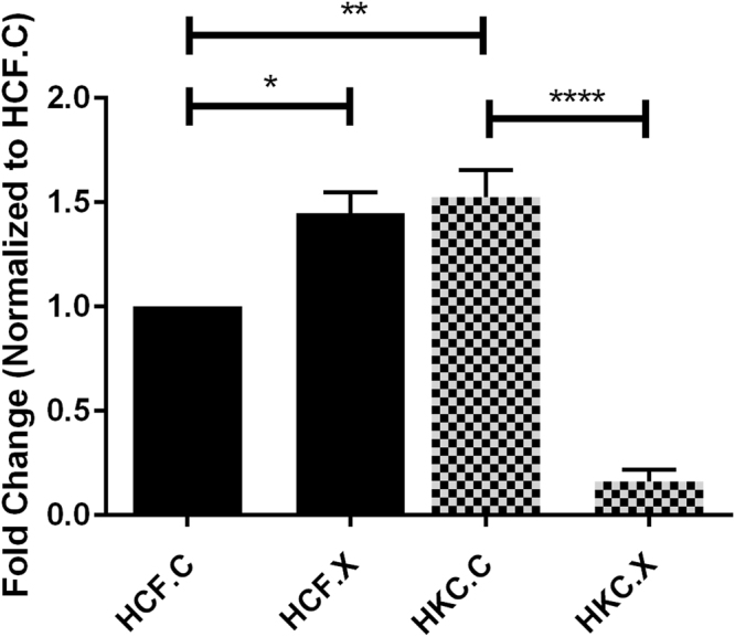
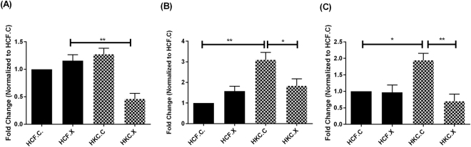
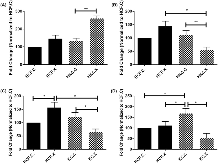
Keratoconus (KC) is a corneal thinning disorder that leads to severe vision impairment As opposed to corneal transplantation; corneal collagen crosslinking (CXL) is a relatively non-invasive procedure that leads to an increase in corneal stiffness. In order to evaluate the effect of CXL on human corneal stromal cells in vitro, we developed a 3-D in vitro CXL model, using primary Human corneal fibroblasts (HCFs) from healthy patients and Human Keratoconus fibroblasts (HKCs) from KC patients. Cells were plated on transwell polycarbonate membranes and stimulated by a stable vitamin C. CXL was performed using a mixed riboflavin 0.1% PBS solution followed by UVA irradiation. Our data revealed no significant apoptosis in either HCFs or HKCs following CXL. However, corneal fibrosis markers, Collagen III and α-smooth muscle actin, were significantly downregulated in CXL HKCs. Furthermore, a significant downregulation was seen in SMAD3, SMAD7, and phosphorylated SMADs -2 and -3 expression in CXL HKCs, contrary to a significant upregulation in both SMAD2 and Lysyl oxidase expression, compared to HCFs. Our novel 3-D in vitro model can be utilized to determine the cellular and molecular effects on the human corneal stroma post CXL, and promises to establish optimized treatment modalities in patients with KC.
圆锥角膜(KC)是一种导致严重视力损害的角膜变薄疾病。与角膜移植相比,角膜胶原交联(CXL)是一种相对非侵入性的程序,可增加角膜硬度。为了评估 CXL 对体外人角膜基质细胞的影响,我们开发了一种 3-D 体外 CXL 模型,使用来自健康患者的原代人角膜成纤维细胞(HCF)和来自 KC 患者的人圆锥角膜成纤维细胞(HKC)。细胞接种在 Transwell 聚碳酸酯膜上,并通过稳定的维生素 C 刺激。CXL 使用混合核黄素 0.1% PBS 溶液进行,然后进行 UVA 照射。我们的数据显示,CXL 后 HCF 或 HKCs 均无明显细胞凋亡。然而,CXL HKCs 中的角膜纤维化标志物 Collagen III 和 α-平滑肌肌动蛋白明显下调。此外,与 HCF 相比,CXL HKCs 中 SMAD3、SMAD7 和磷酸化 SMADs-2 和 -3 的表达明显下调,而 SMAD2 和赖氨酰氧化酶的表达明显上调。我们的新型 3-D 体外模型可用于确定 CXL 后对人角膜基质的细胞和分子影响,并有望为 KC 患者建立优化的治疗方式。