Adult Stem Cell Section, National Institute of Dental and Craniofacial Research, National Institutes of Health, Bethesda, MD 20892;
Adult Stem Cell Section, National Institute of Dental and Craniofacial Research, National Institutes of Health, Bethesda, MD 20892.
Proc Natl Acad Sci U S A. 2021 Jan 19;118(3). doi: 10.1073/pnas.2002574118.
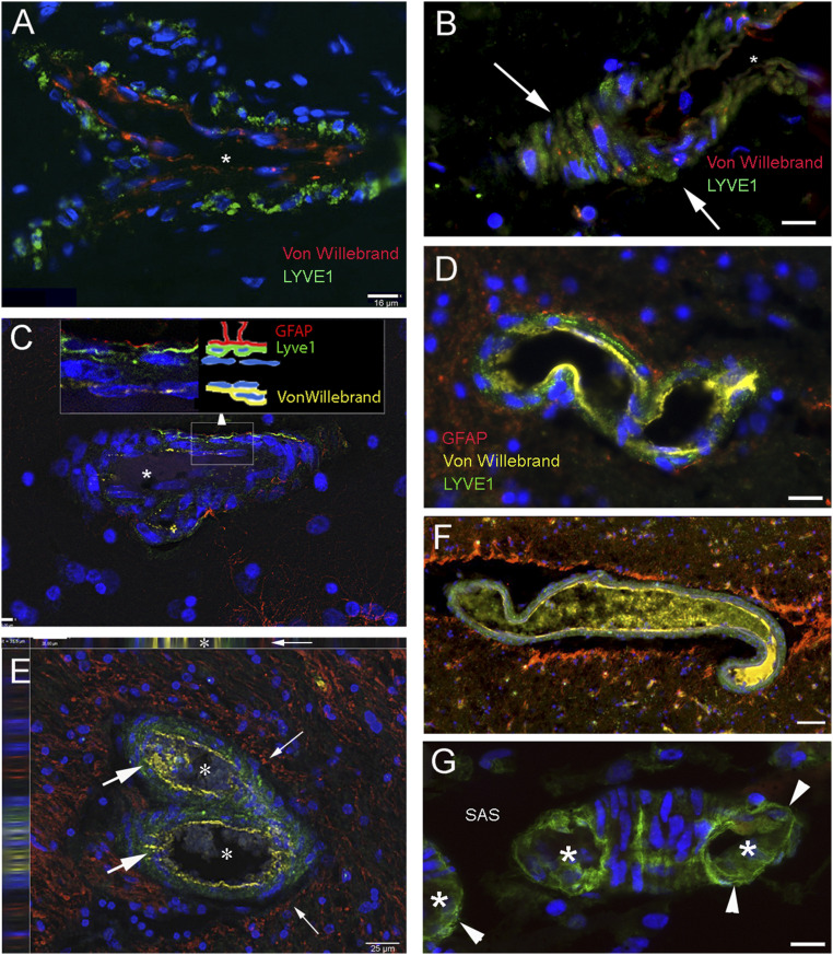
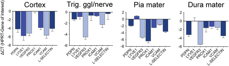
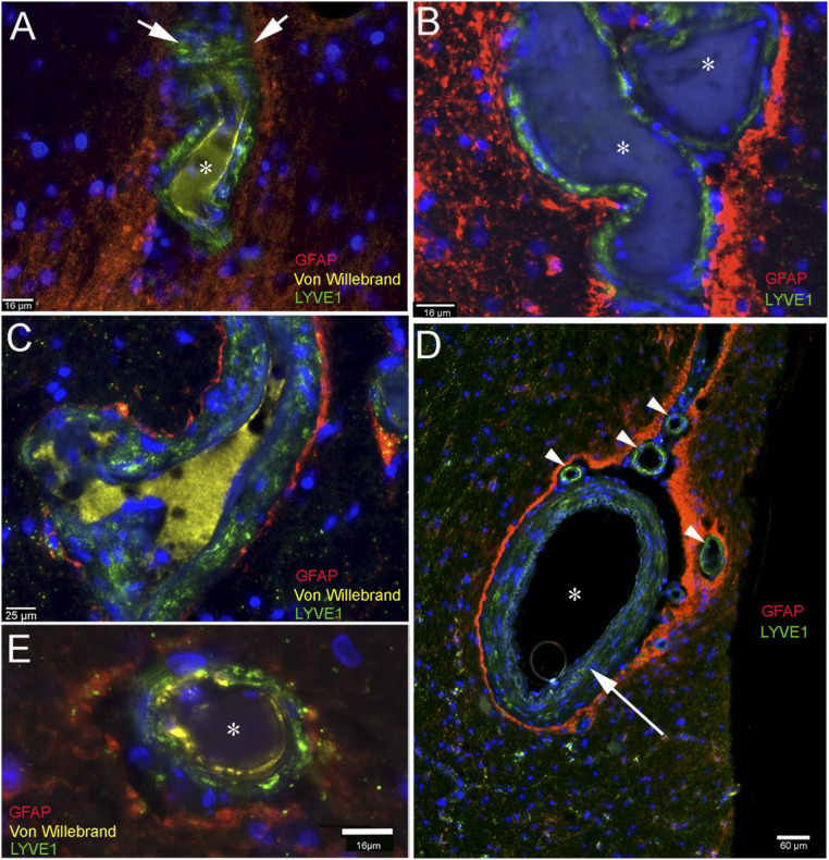
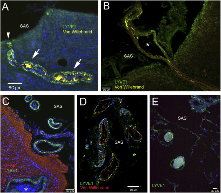
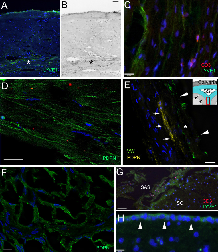
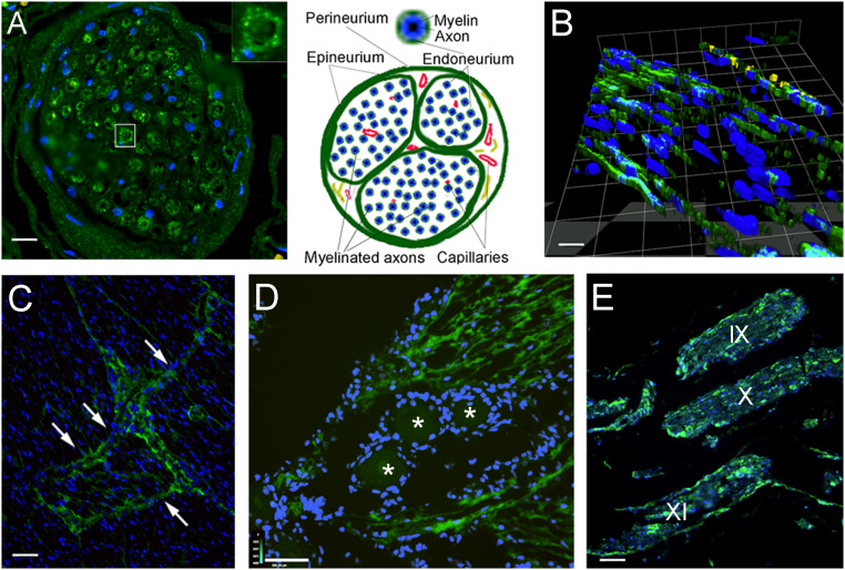
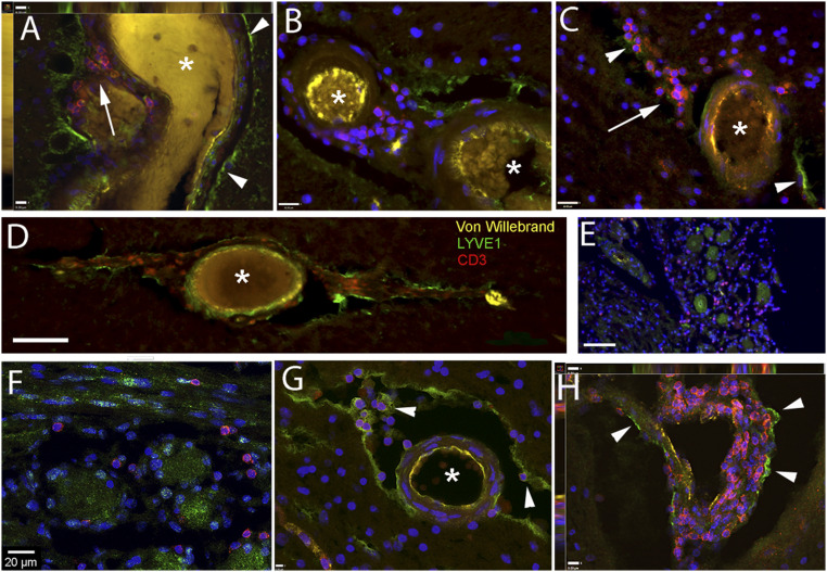
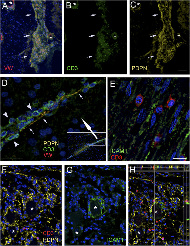
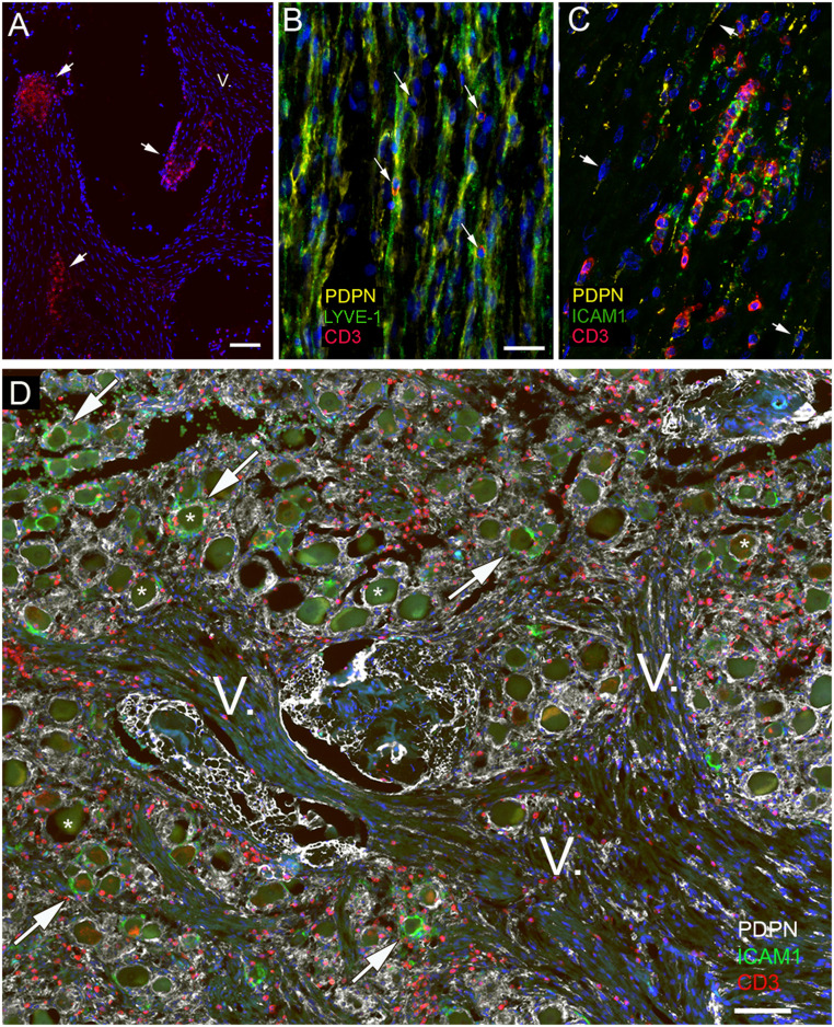
Almost 150 papers about brain lymphatics have been published in the last 150 years. Recently, the information in these papers has been synthesized into a picture of central nervous system (CNS) "glymphatics," but the fine structure of lymphatic elements in the human brain based on imaging specific markers of lymphatic endothelium has not been described. We used LYVE1 and PDPN antibodies to visualize lymphatic marker-positive cells (LMPCs) in postmortem human brain samples, meninges, cavernous sinus (cavum trigeminale), and cranial nerves and bolstered our findings with a VEGFR3 antibody. LMPCs were present in the perivascular space, the walls of small and large arteries and veins, the media of large vessels along smooth muscle cell membranes, and the vascular adventitia. Lymphatic marker staining was detected in the pia mater, in the arachnoid, in venous sinuses, and among the layers of the dura mater. There were many LMPCs in the perineurium and endoneurium of cranial nerves. Soluble waste may move from the brain parenchyma via perivascular and paravascular routes to the closest subarachnoid space and then travel along the dura mater and/or cranial nerves. Particulate waste products travel along the laminae of the dura mater toward the jugular fossa, lamina cribrosa, and perineurium of the cranial nerves to enter the cervical lymphatics. CD3-positive T cells appear to be in close proximity to LMPCs in perivascular/perineural spaces throughout the brain. Both immunostaining and qPCR confirmed the presence of adhesion molecules in the CNS known to be involved in T cell migration.
在过去的 150 年中,已经发表了将近 150 篇关于脑淋巴的论文。最近,这些论文中的信息被综合成中枢神经系统(CNS)“神经淋巴学”的图片,但尚未描述基于成像淋巴内皮特定标志物的人脑中淋巴样元素的精细结构。我们使用 LYVE1 和 PDPN 抗体来可视化尸检人脑样本、脑膜、海绵窦(三叉神经腔)和颅神经中的淋巴标志物阳性细胞(LMPC),并使用 VEGFR3 抗体来支持我们的发现。LMPC 存在于血管周围间隙、小动脉和静脉的壁、大血管的中膜以及平滑肌细胞膜的血管外膜中。在软膜、蛛网膜、静脉窦和硬脑膜的层间都可以检测到淋巴标志物染色。许多 LMPC 存在于颅神经的神经外膜和神经内膜中。可溶性废物可能通过血管周围和血管旁途径从脑实质转移到最近的蛛网膜下腔,然后沿硬脑膜和/或颅神经移动。颗粒状废物沿着硬脑膜的层面向颈静脉窝、筛板和颅神经的神经外膜移动,进入颈淋巴。CD3 阳性 T 细胞似乎与整个大脑血管周围/神经周围空间中的 LMPC 密切相关。免疫染色和 qPCR 都证实了 CNS 中参与 T 细胞迁移的黏附分子的存在。