Department of Pathology, Faculty of Medicine, Comenius University, Sasinkova 4, 811 08, Bratislava, Slovakia.
Department of Pathology, Faculty Hospital, A. Zarnova 11, 917 75, Trnava, Slovakia.
Sci Rep. 2021 Jan 14;11(1):1294. doi: 10.1038/s41598-020-80351-9.
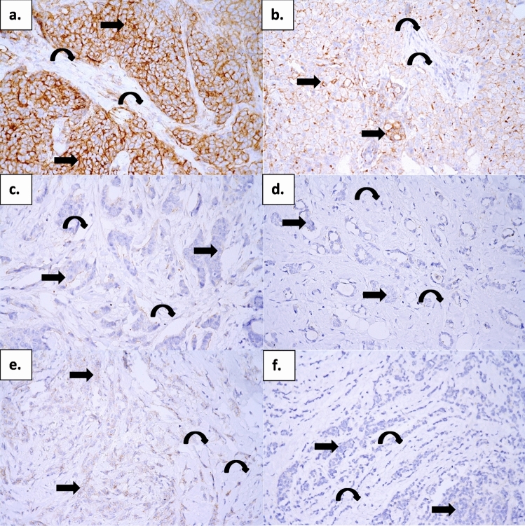
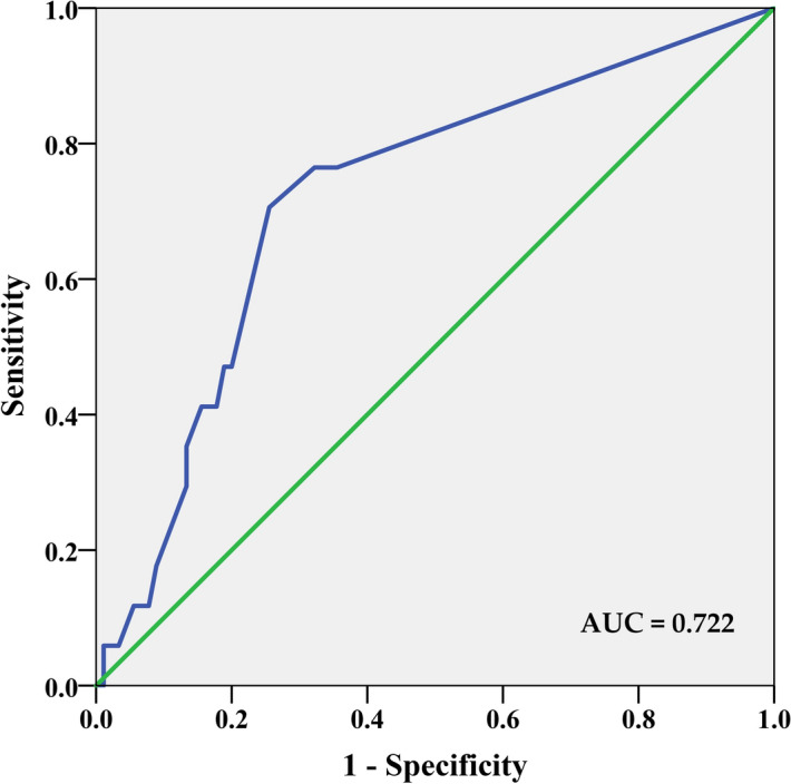
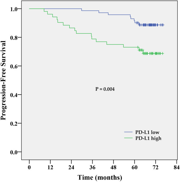
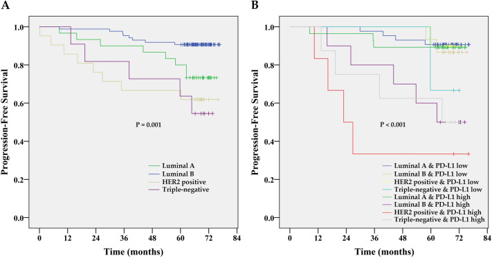
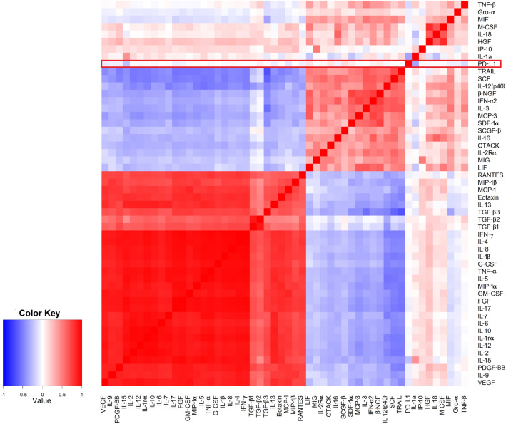
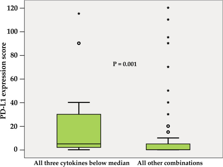
Programmed death ligand 1 (PD-L1) overexpression has been associated with poor clinical outcomes in several human cancers whose increased malignant behaviour might be related to PD-L1 mediated systemic immunological tolerance. This study aims to verify if circulating cytokines may serve as a proxy for non-invasive identification of sensitive prognostic biomarkers reflecting tumour and its microenvironment. Immunohistochemistry was used to measure PD-L1 expression in tumour tissue sections of 148 chemonaïve breast cancer (BC) patients. The panel of 51 cytokines was analysed using multiplex bead arrays. High PD-L1 expression in tumours was associated with shorter progression-free survival (HR 3.25; 95% CI 1.39-7.61; P = 0.006) and low circulating levels of three multifunctional molecules; VEGF, TNF-β and IL-15 (P = 0.001). In multivariate analysis, patients with low VEGF had 4.6-fold increased risk of PD-L1 overexpression (P = 0.008), present in 76.5% of patients with all these three cytokines below the median (vs. 35.6% among the others; P = 0.002). The area under the curve value of 0.722 (95% CI 0.59-0.85; P = 0.004) shows that this combination of cytokines has a moderate ability to discriminate between PD-L1 high vs. PD-L1 low patients. Plasma cytokines, therefore, could serve as potential non-invasive biomarkers for the identification of high-risk BC cases.
程序性死亡配体 1(PD-L1)过表达与多种人类癌症的不良临床结局相关,其恶性行为增加可能与 PD-L1 介导的全身免疫耐受有关。本研究旨在验证循环细胞因子是否可作为替代物,用于非侵入性识别反映肿瘤及其微环境的敏感预后生物标志物。免疫组织化学用于测量 148 例未经化疗的乳腺癌(BC)患者肿瘤组织切片中的 PD-L1 表达。使用多重珠阵列分析了 51 种细胞因子的组合。肿瘤中 PD-L1 高表达与无进展生存期较短相关(HR 3.25;95%CI 1.39-7.61;P=0.006),并且循环水平低的三种多功能分子;VEGF、TNF-β 和 IL-15(P=0.001)。在多变量分析中,低 VEGF 的患者 PD-L1 过表达的风险增加了 4.6 倍(P=0.008),在所有这三种细胞因子均低于中位数的患者中,存在 76.5%(而在其他患者中存在 35.6%;P=0.002)。曲线下面积为 0.722(95%CI 0.59-0.85;P=0.004),表明该细胞因子组合具有中等能力来区分 PD-L1 高与 PD-L1 低患者。因此,血浆细胞因子可作为识别高危 BC 病例的潜在非侵入性生物标志物。