• 文献检索
  • 文档翻译
  • 深度研究
  • 学术资讯
  • Suppr Zotero 插件Zotero 插件
  • 邀请有礼
  • 套餐&价格
  • 历史记录
应用&插件
Suppr Zotero 插件Zotero 插件浏览器插件Mac 客户端Windows 客户端微信小程序
定价
高级版会员购买积分包购买API积分包
服务
文献检索文档翻译深度研究API 文档MCP 服务
关于我们
关于 Suppr公司介绍联系我们用户协议隐私条款
关注我们

Suppr 超能文献

核心技术专利:CN118964589B侵权必究
粤ICP备2023148730 号-1Suppr @ 2026

文献检索

告别复杂PubMed语法,用中文像聊天一样搜索,搜遍4000万医学文献。AI智能推荐,让科研检索更轻松。

立即免费搜索

文件翻译

保留排版,准确专业,支持PDF/Word/PPT等文件格式,支持 12+语言互译。

免费翻译文档

深度研究

AI帮你快速写综述,25分钟生成高质量综述,智能提取关键信息,辅助科研写作。

立即免费体验

血吸虫病假瘤型酷似神经内分泌肿瘤:病例报告并简要复习腹膜后肿块的鉴别诊断。

Pseudotumoral form of schistosomiasis mimicking neuroendocrine tumor: a case report and brief review of the differential diagnosis of retroperitoneal masses.

机构信息

Sección de Endocrinología y Nutrición, Hospital General Universitario Santa Lucía, Cartagena, Spain.

Servicio de Anatomía Patológica, Hospital General Universitario Santa Lucía, Cartagena, Spain.

出版信息

Pan Afr Med J. 2020 Oct 27;37:186. doi: 10.11604/pamj.2020.37.186.26344. eCollection 2020.

DOI:10.11604/pamj.2020.37.186.26344
PMID:33447341
原文链接:https://pmc.ncbi.nlm.nih.gov/articles/PMC7778187/
Abstract

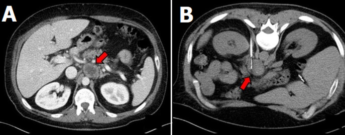
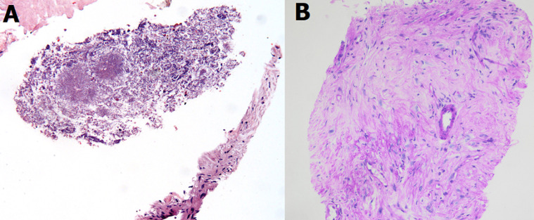
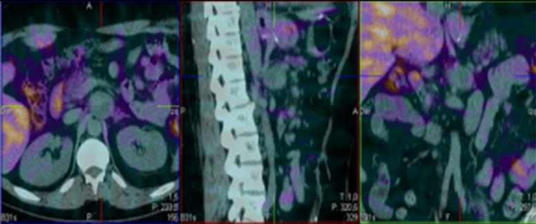
Differential diagnosis of retroperitoneal masses may become complex and requires careful anamnesis, physical examination and several complementary tests. We present the clinical case of a male patient aged 45 years who was diagnosed with a 4cm paraaortic lesion compatible with neuroendocrine tumor in the abdominal computed tomography (CT) exam. The workup performed with SPECT-CT, somatostatin receptors scintigraphy, MIBG scintigraphy, 24-hour urine total and fractionated catecholamines and 24-hour urine 5-OH indoleacetic did not confirm the first diagnostic impression. Finally, the lesion was biopsied and presence of micro-organisms was revealed. Further exams confirmed schistosomiasis as the cause of the paraaortic lesion. Histological diagnosis can be helpful with regard to the differential diagnosis of retroperitoneal masses.

摘要

腹膜后肿块的鉴别诊断可能变得复杂,需要仔细的病史询问、体格检查和几项补充检查。我们呈现了一位 45 岁男性患者的临床病例,其在腹部计算机断层扫描(CT)检查中被诊断为 4cm 主动脉旁病变,符合神经内分泌肿瘤。通过 SPECT-CT、生长抑素受体闪烁显像、MIBG 闪烁显像、24 小时尿总儿茶酚胺和分馏儿茶酚胺、24 小时尿 5-羟吲哚乙酸进行的检查未证实最初的诊断印象。最后,对病变进行了活检,发现了微生物。进一步的检查证实了该患者患有血吸虫病,这是导致主动脉旁病变的原因。组织学诊断对于腹膜后肿块的鉴别诊断可能有帮助。

https://cdn.ncbi.nlm.nih.gov/pmc/blobs/b246/7778187/004fd792c2b7/PAMJ-37-186-g003.jpg
https://cdn.ncbi.nlm.nih.gov/pmc/blobs/b246/7778187/bbadd8182b3e/PAMJ-37-186-g001.jpg
https://cdn.ncbi.nlm.nih.gov/pmc/blobs/b246/7778187/8a647908133b/PAMJ-37-186-g002.jpg
https://cdn.ncbi.nlm.nih.gov/pmc/blobs/b246/7778187/004fd792c2b7/PAMJ-37-186-g003.jpg
https://cdn.ncbi.nlm.nih.gov/pmc/blobs/b246/7778187/bbadd8182b3e/PAMJ-37-186-g001.jpg
https://cdn.ncbi.nlm.nih.gov/pmc/blobs/b246/7778187/8a647908133b/PAMJ-37-186-g002.jpg
https://cdn.ncbi.nlm.nih.gov/pmc/blobs/b246/7778187/004fd792c2b7/PAMJ-37-186-g003.jpg

相似文献

1
Pseudotumoral form of schistosomiasis mimicking neuroendocrine tumor: a case report and brief review of the differential diagnosis of retroperitoneal masses.血吸虫病假瘤型酷似神经内分泌肿瘤:病例报告并简要复习腹膜后肿块的鉴别诊断。
Pan Afr Med J. 2020 Oct 27;37:186. doi: 10.11604/pamj.2020.37.186.26344. eCollection 2020.
2
Primary cystic and solid neuroendocrine tumor of the retroperitoneum: A case report.腹膜后原发性囊实性神经内分泌肿瘤:一例报告。
Medicine (Baltimore). 2021 Jan 15;100(2):e24054. doi: 10.1097/MD.0000000000024054.
3
Primary Epithelial Neuroendocrine Tumors of the Retroperitoneum.原发性腹膜后上皮性神经内分泌肿瘤
Perm J. 2015 Fall;19(4):71-5. doi: 10.7812/TPP/15-058.
4
[Pseudotumoral retroperitoneal tuberculosis. Apropos of a case].
J Radiol. 1986 Feb;67(2):141-3.
5
Spontaneous retroperitoneal abscess as the first clinical manifestation of a non-functioning retroperitoneal paraganglioma.自发性腹膜后脓肿作为无功能腹膜后副神经节瘤的首发临床表现。
Ann R Coll Surg Engl. 2010 Apr;92(3):W17-9. doi: 10.1308/147870810X12659688851555.
6
Management dilemmas of retroperitoneal paraganglioma: report of a case.腹膜后副神经节瘤的管理困境:一例报告
Rev Med Chir Soc Med Nat Iasi. 2014 Jul-Sep;118(3):679-82.
7
Primary retroperitoneal masses: what is the differential diagnosis?原发性腹膜后肿块:鉴别诊断有哪些?
Abdom Imaging. 2015 Aug;40(6):1887-903. doi: 10.1007/s00261-014-0311-x.
8
[Case of retroperitoneal paraganglioma].[腹膜后副神经节瘤病例]
Hinyokika Kiyo. 2007 Nov;53(11):801-4.
9
Incremental value of Tc-HYNIC-TOC SPECT/CT over whole-body planar scintigraphy and SPECT in patients with neuroendocrine tumours.锝[Tc] 泮替膦酸盐单光子发射计算机断层显像/计算机断层扫描(Tc-HYNIC-TOC SPECT/CT)相对于全身平面闪烁显像和单光子发射计算机断层扫描(SPECT)在神经内分泌肿瘤患者中的增量价值
Nuklearmedizin. 2017 Jun 12;56(3):97-107. doi: 10.3413/Nukmed-0851-16-10. Epub 2017 Feb 6.
10
A case of retroperitoneal extramedullary plasmacytoma with multiple metastases.一例伴有多处转移的腹膜后髓外浆细胞瘤。
Clin Imaging. 2000 Nov-Dec;24(6):365-7. doi: 10.1016/s0899-7071(00)00247-3.

引用本文的文献

1
A case of Schistosoma japonicum retroperitoneal pseudotumor diagnosed by cone-beam CT-guided coaxial biopsy system.1例通过锥形束CT引导同轴活检系统诊断的日本血吸虫腹膜后假性肿瘤病例。
Acta Radiol Open. 2022 Sep 24;11(9):20584601221129153. doi: 10.1177/20584601221129153. eCollection 2022 Sep.
2
Huge Colonic Granuloma of Schistosomiasis Mimicked Cancer in a 10-Years-Old Child: A Case Report.一名10岁儿童的血吸虫病巨大结肠肉芽肿酷似癌症:病例报告
Int Med Case Rep J. 2022 Jul 21;15:379-384. doi: 10.2147/IMCRJ.S372186. eCollection 2022.
3
Giant Retroperitoneal Malignant Peripheral Nerve Sheath Tumor Treated with Multiorgan Resection: a Case Report and Review of the Literature.

本文引用的文献

1
Pheochromocytoma and Paraganglioma.嗜铬细胞瘤和副神经节瘤。
N Engl J Med. 2019 Aug 8;381(6):552-565. doi: 10.1056/NEJMra1806651.
2
Schistosomiasis: Life Cycle, Diagnosis, and Control.血吸虫病:生命周期、诊断与控制
Curr Ther Res Clin Exp. 2019 Jun 22;91:5-9. doi: 10.1016/j.curtheres.2019.06.001. eCollection 2019.
3
Schistosome Egg Migration: Mechanisms, Pathogenesis and Host Immune Responses.血吸虫卵迁移:机制、发病机制和宿主免疫反应。
多器官切除治疗巨大腹膜后恶性周围神经鞘瘤:病例报告及文献复习
Indian J Surg Oncol. 2022 Jun;13(2):372-376. doi: 10.1007/s13193-021-01477-4. Epub 2021 Nov 8.
4
Medical Imaging in the Diagnosis of Schistosomiasis: A Review.医学成像在血吸虫病诊断中的应用综述
Pathogens. 2021 Aug 20;10(8):1058. doi: 10.3390/pathogens10081058.
Front Immunol. 2018 Dec 20;9:3042. doi: 10.3389/fimmu.2018.03042. eCollection 2018.
4
Practical approach to primary retroperitoneal masses in adults.成人原发性腹膜后肿块的实用处理方法。
Radiol Bras. 2018 Nov-Dec;51(6):391-400. doi: 10.1590/0100-3984.2017.0179.
5
Magnetic Resonance Imaging of the Perirenal Space and Retroperitoneum.
Magn Reson Imaging Clin N Am. 2019 Feb;27(1):77-103. doi: 10.1016/j.mric.2018.08.007. Epub 2018 Oct 29.
6
Schistosoma "Eggs-Iting" the Host: Granuloma Formation and Egg Excretion.血吸虫“鸡蛋-激动人心”宿主:肉芽肿形成和卵排泄。
Front Immunol. 2018 Oct 29;9:2492. doi: 10.3389/fimmu.2018.02492. eCollection 2018.
7
Retroperitoneal paraganglioma: a chameleon masquerading as an adrenal pheochromocytoma.腹膜后副神经节瘤:伪装成肾上腺嗜铬细胞瘤的变色龙。
Ann R Coll Surg Engl. 2019 Feb;101(2):e62-e65. doi: 10.1308/rcsann.2018.0193. Epub 2018 Oct 29.
8
Current status of functional imaging in neuroblastoma, pheochromocytoma, and paraganglioma disease.神经母细胞瘤、嗜铬细胞瘤和副神经节瘤疾病中功能成像的现状。
Wien Med Wochenschr. 2019 Feb;169(1-2):25-32. doi: 10.1007/s10354-018-0658-7. Epub 2018 Sep 4.
9
Schistosomiasis.血吸虫病。
Nat Rev Dis Primers. 2018 Aug 9;4(1):13. doi: 10.1038/s41572-018-0013-8.
10
A retroperitoneal mass needs respect.腹膜后肿块需要引起重视。
ANZ J Surg. 2018 Apr;88(4):261-262. doi: 10.1111/ans.14354.