• 文献检索
  • 文档翻译
  • 深度研究
  • 学术资讯
  • Suppr Zotero 插件Zotero 插件
  • 邀请有礼
  • 套餐&价格
  • 历史记录
应用&插件
Suppr Zotero 插件Zotero 插件浏览器插件Mac 客户端Windows 客户端微信小程序
定价
高级版会员购买积分包购买API积分包
服务
文献检索文档翻译深度研究API 文档MCP 服务
关于我们
关于 Suppr公司介绍联系我们用户协议隐私条款
关注我们

Suppr 超能文献

核心技术专利:CN118964589B侵权必究
粤ICP备2023148730 号-1Suppr @ 2026

文献检索

告别复杂PubMed语法,用中文像聊天一样搜索,搜遍4000万医学文献。AI智能推荐,让科研检索更轻松。

立即免费搜索

文件翻译

保留排版,准确专业,支持PDF/Word/PPT等文件格式,支持 12+语言互译。

免费翻译文档

深度研究

AI帮你快速写综述,25分钟生成高质量综述,智能提取关键信息,辅助科研写作。

立即免费体验

检测创伤性脑损伤后个体患者的轴索损伤。

Detecting axonal injury in individual patients after traumatic brain injury.

机构信息

Clinical, cognitive and computational neuroimaging laboratory (C3NL), Department of Brain Sciences, Faculty of Medicine, Imperial College London, London, W12 0NN, UK.

UK Dementia Research Institute Care Research and Technology Centre, Imperial College London and the University of Surrey, London, W12 0NN UK.

出版信息

Brain. 2021 Feb 12;144(1):92-113. doi: 10.1093/brain/awaa372.

DOI:10.1093/brain/awaa372
PMID:33257929
原文链接:https://pmc.ncbi.nlm.nih.gov/articles/PMC7880666/
Abstract

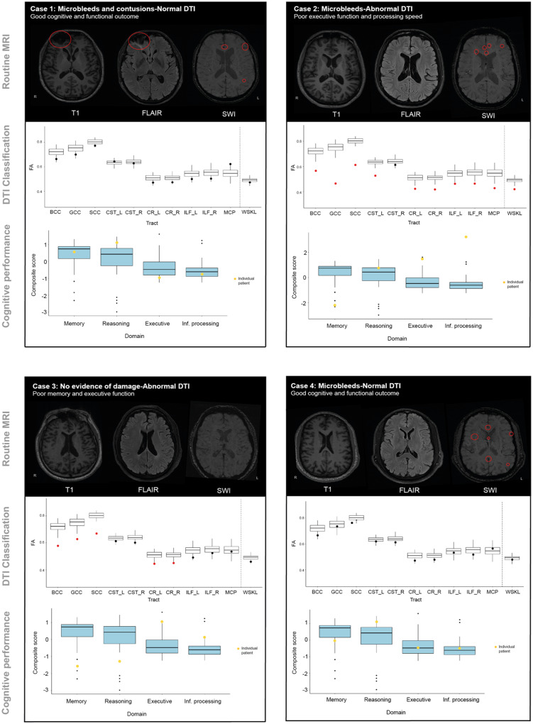
Poor outcomes after traumatic brain injury (TBI) are common yet remain difficult to predict. Diffuse axonal injury is important for outcomes, but its assessment remains limited in the clinical setting. Currently, axonal injury is diagnosed based on clinical presentation, visible damage to the white matter or via surrogate markers of axonal injury such as microbleeds. These do not accurately quantify axonal injury leading to misdiagnosis in a proportion of patients. Diffusion tensor imaging provides a quantitative measure of axonal injury in vivo, with fractional anisotropy often used as a proxy for white matter damage. Diffusion imaging has been widely used in TBI but is not routinely applied clinically. This is in part because robust analysis methods to diagnose axonal injury at the individual level have not yet been developed. Here, we present a pipeline for diffusion imaging analysis designed to accurately assess the presence of axonal injury in large white matter tracts in individuals. Average fractional anisotropy is calculated from tracts selected on the basis of high test-retest reliability, good anatomical coverage and their association to cognitive and clinical impairments after TBI. We test our pipeline for common methodological issues such as the impact of varying control sample sizes, focal lesions and age-related changes to demonstrate high specificity, sensitivity and test-retest reliability. We assess 92 patients with moderate-severe TBI in the chronic phase (≥6 months post-injury), 25 patients in the subacute phase (10 days to 6 weeks post-injury) with 6-month follow-up and a large control cohort (n = 103). Evidence of axonal injury is identified in 52% of chronic and 28% of subacute patients. Those classified with axonal injury had significantly poorer cognitive and functional outcomes than those without, a difference not seen for focal lesions or microbleeds. Almost a third of patients with unremarkable standard MRIs had evidence of axonal injury, whilst 40% of patients with visible microbleeds had no diffusion evidence of axonal injury. More diffusion abnormality was seen with greater time since injury, across individuals at various chronic injury times and within individuals between subacute and 6-month scans. We provide evidence that this pipeline can be used to diagnose axonal injury in individual patients at subacute and chronic time points, and that diffusion MRI provides a sensitive and complementary measure when compared to susceptibility weighted imaging, which measures diffuse vascular injury. Guidelines for the implementation of this pipeline in a clinical setting are discussed.

摘要

创伤性脑损伤 (TBI) 后预后不良较为常见,但仍难以预测。弥漫性轴索损伤对预后很重要,但在临床环境中其评估仍受到限制。目前,轴索损伤是基于临床表现、白质的可见损伤或轴索损伤的替代标志物(如微出血)来诊断的。这些方法并不能准确地量化轴索损伤,导致一部分患者误诊。弥散张量成像可提供体内轴索损伤的定量测量,各向异性分数常被用作白质损伤的替代物。弥散成像已广泛应用于 TBI 中,但并未常规应用于临床。这在一定程度上是因为尚未开发出用于个体水平诊断轴索损伤的稳健分析方法。在这里,我们提出了一种弥散成像分析的工作流程,旨在准确评估个体中大的白质束中轴索损伤的存在。基于高测试-重测可靠性、良好的解剖学覆盖范围以及它们与 TBI 后认知和临床障碍的相关性,选择轨迹来计算平均各向异性分数。我们测试了我们的工作流程是否存在常见的方法学问题,如不同对照组样本量、局灶性病变和与年龄相关的变化的影响,以证明其具有高特异性、敏感性和测试-重测可靠性。我们评估了 92 名处于慢性期(受伤后≥6 个月)的中重度 TBI 患者、25 名亚急性期(受伤后 10 天至 6 周)患者和一个大的对照组(n=103),并进行了 6 个月的随访。在慢性期和亚急性期患者中分别有 52%和 28%的患者发现了轴索损伤的证据。与没有轴索损伤的患者相比,被分类为有轴索损伤的患者认知和功能结局明显更差,而局灶性病变或微出血的患者则没有这种差异。将近三分之一的 MRI 未见明显异常的患者有轴索损伤的证据,而 40%有可见微出血的患者则没有弥散异常的证据。随着时间的推移,个体在不同的慢性损伤时间点以及个体在亚急性期和 6 个月扫描之间,弥散异常的情况更明显。我们提供的证据表明,该工作流程可用于在亚急性期和慢性期诊断个体患者的轴索损伤,与测量弥漫性血管损伤的磁敏感加权成像相比,弥散 MRI 是一种更敏感和互补的测量方法。讨论了在临床环境中实施该工作流程的指南。

https://cdn.ncbi.nlm.nih.gov/pmc/blobs/019c/7880666/98f1f83e1ebe/awaa372f8.jpg
https://cdn.ncbi.nlm.nih.gov/pmc/blobs/019c/7880666/cb1265f704c0/awaa372f1.jpg
https://cdn.ncbi.nlm.nih.gov/pmc/blobs/019c/7880666/87dc76ef3859/awaa372f2.jpg
https://cdn.ncbi.nlm.nih.gov/pmc/blobs/019c/7880666/8097ad20d6e7/awaa372f3.jpg
https://cdn.ncbi.nlm.nih.gov/pmc/blobs/019c/7880666/66dd8f094d54/awaa372f4.jpg
https://cdn.ncbi.nlm.nih.gov/pmc/blobs/019c/7880666/ef586f3c7aee/awaa372f5.jpg
https://cdn.ncbi.nlm.nih.gov/pmc/blobs/019c/7880666/a25e44958ac6/awaa372f6.jpg
https://cdn.ncbi.nlm.nih.gov/pmc/blobs/019c/7880666/7850bcc4e405/awaa372f7.jpg
https://cdn.ncbi.nlm.nih.gov/pmc/blobs/019c/7880666/98f1f83e1ebe/awaa372f8.jpg

相似文献

1
Detecting axonal injury in individual patients after traumatic brain injury.检测创伤性脑损伤后个体患者的轴索损伤。
Brain. 2021 Feb 12;144(1):92-113. doi: 10.1093/brain/awaa372.
2
Diffuse axonal injury predicts neurodegeneration after moderate-severe traumatic brain injury.弥漫性轴索损伤预测中重度创伤性脑损伤后的神经退行性变。
Brain. 2020 Dec 1;143(12):3685-3698. doi: 10.1093/brain/awaa316.
3
Limited Colocalization of Microbleeds and Microstructural Changes after Severe Traumatic Brain Injury.严重创伤性脑损伤后微出血与微观结构变化的局限性共存。
J Neurotrauma. 2020 Feb 15;37(4):581-592. doi: 10.1089/neu.2019.6608. Epub 2019 Nov 20.
4
Automated detection of axonal damage along white matter tracts in acute severe traumatic brain injury.自动检测急性严重创伤性脑损伤中沿白质束的轴突损伤。
Neuroimage Clin. 2023;37:103294. doi: 10.1016/j.nicl.2022.103294. Epub 2022 Dec 13.
5
Traumatic axonal injury: is the prognostic information produced by conventional MRI and DTI complementary or supplementary?创伤性轴索损伤:常规 MRI 和 DTI 提供的预后信息是互补的还是补充的?
J Neurosurg. 2021 Jul 2;136(1):242-256. doi: 10.3171/2020.11.JNS203124. Print 2022 Jan 1.
6
From biomechanics to pathology: predicting axonal injury from patterns of strain after traumatic brain injury.从生物力学到病理学:从创伤性脑损伤后的应变模式预测轴突损伤。
Brain. 2021 Feb 12;144(1):70-91. doi: 10.1093/brain/awaa336.
7
Axonal disruption in white matter underlying cortical sulcus tau pathology in chronic traumatic encephalopathy.皮质沟回tau 病理学下白质中轴索中断与慢性创伤性脑病有关。
Acta Neuropathol. 2017 Mar;133(3):367-380. doi: 10.1007/s00401-017-1686-x. Epub 2017 Feb 18.
8
Common Patterns of Regional Brain Injury Detectable by Diffusion Tensor Imaging in Otherwise Normal-Appearing White Matter in Patients with Early Moderate to Severe Traumatic Brain Injury.早期中重度创伤性脑损伤患者,弥散张量成像可发现正常外观的脑白质中存在的常见区域性脑损伤模式。
J Neurotrauma. 2018 Mar 1;35(5):739-749. doi: 10.1089/neu.2016.4944. Epub 2018 Feb 9.
9
Traumatic axonal injury: Relationships between lesions in the early phase and diffusion tensor imaging parameters in the chronic phase of traumatic brain injury.创伤性轴索损伤:创伤性脑损伤慢性期早期病变与扩散张量成像参数之间的关系
J Neurosci Res. 2016 Jul;94(7):623-35. doi: 10.1002/jnr.23728. Epub 2016 Mar 6.
10
Imaging Markers for the Characterization of Gray and White Matter Changes from Acute to Chronic Stages after Experimental Traumatic Brain Injury.实验性颅脑损伤后从急性期到慢性期的灰、白质变化的影像学标志物。
J Neurotrauma. 2021 Jun 15;38(12):1642-1653. doi: 10.1089/neu.2020.7151. Epub 2021 Jan 11.

引用本文的文献

1
Structural injuries are associated with better emotional wellbeing after traumatic brain injury.创伤性脑损伤后,结构性损伤与更好的情绪健康状况相关。
medRxiv. 2025 Jul 25:2025.07.24.25332156. doi: 10.1101/2025.07.24.25332156.
2
White matter tract differences in persistent post-traumatic headache, migraine, and healthy controls: a diffusion tensor imaging study.创伤后持续性头痛、偏头痛与健康对照者的白质束差异:一项扩散张量成像研究
J Headache Pain. 2025 Jul 4;26(1):155. doi: 10.1186/s10194-025-02084-2.
3
Meso-scale network analysis of resting state-fMRI brain network connectivity performs poorly as a prognostic tool in critically ill traumatic brain injury patients.

本文引用的文献

1
Traumatic microbleeds suggest vascular injury and predict disability in traumatic brain injury.外伤性微出血提示血管损伤,并可预测外伤性脑损伤的残疾。
Brain. 2019 Nov 1;142(11):3550-3564. doi: 10.1093/brain/awz290.
2
Understanding neurodegeneration after traumatic brain injury: from mechanisms to clinical trials in dementia.创伤性脑损伤后神经退行性变的理解:从机制到痴呆症的临床试验。
J Neurol Neurosurg Psychiatry. 2019 Nov;90(11):1221-1233. doi: 10.1136/jnnp-2017-317557. Epub 2019 Sep 21.
3
Retrospective harmonization of multi-site diffusion MRI data acquired with different acquisition parameters.
作为重症创伤性脑损伤患者的预后工具,静息态功能磁共振成像脑网络连通性的中尺度网络分析表现不佳。
Neuroimage Rep. 2022 Jan 10;2(1):100079. doi: 10.1016/j.ynirp.2022.100079. eCollection 2022 Mar.
4
Revisiting the critical roles of reactive microglia in traumatic brain injury.重新审视反应性小胶质细胞在创伤性脑损伤中的关键作用。
Int J Surg. 2025 Jun 1;111(6):3942-3978. doi: 10.1097/JS9.0000000000002420. Epub 2025 May 12.
5
Mild traumatic brain injury increases cortical iron: evidence from individual susceptibility mapping.轻度创伤性脑损伤会增加皮质铁含量:来自个体易感性图谱的证据。
Brain Commun. 2025 Mar 12;7(2):fcaf110. doi: 10.1093/braincomms/fcaf110. eCollection 2025.
6
Cortical lesions and focal white matter injury are associated with attentional performance in chronic traumatic brain injury.皮质病变和局灶性白质损伤与慢性创伤性脑损伤的注意力表现相关。
Brain Commun. 2024 Nov 21;7(1):fcae420. doi: 10.1093/braincomms/fcae420. eCollection 2025.
7
Diffusion Tensor Imaging as Neurologic Predictor in Patients Affected by Traumatic Brain Injury: Scoping Review.扩散张量成像作为创伤性脑损伤患者的神经学预测指标:范围综述
Brain Sci. 2025 Jan 14;15(1):70. doi: 10.3390/brainsci15010070.
8
Aberrant dynamic functional network connectivity in patients with diffuse axonal injury.弥散性轴索损伤患者异常的动态功能网络连接。
Sci Rep. 2024 Nov 9;14(1):27386. doi: 10.1038/s41598-024-79052-4.
9
High-dimensional proteomic analysis for pathophysiological classification of traumatic brain injury.用于创伤性脑损伤病理生理分类的高维蛋白质组学分析
Brain. 2025 Mar 6;148(3):1015-1030. doi: 10.1093/brain/awae305.
10
Automated Measurement of Cerebral Hemorrhagic Contusions and Outcomes After Traumatic Brain Injury in the TRACK-TBI Study.TRACK-TBI 研究中创伤性脑损伤后脑出血性挫伤的自动测量和结果。
JAMA Netw Open. 2024 Aug 1;7(8):e2427772. doi: 10.1001/jamanetworkopen.2024.27772.
回顾性协调多部位扩散 MRI 数据,这些数据是使用不同采集参数采集的。
Neuroimage. 2019 Jan 1;184:180-200. doi: 10.1016/j.neuroimage.2018.08.073. Epub 2018 Sep 8.
4
Dopaminergic abnormalities following traumatic brain injury.创伤性脑损伤后的多巴胺能异常。
Brain. 2018 Mar 1;141(3):797-810. doi: 10.1093/brain/awx357.
5
Spatial patterns of progressive brain volume loss after moderate-severe traumatic brain injury.中重度创伤性脑损伤后脑容量进行性损失的空间模式。
Brain. 2018 Mar 1;141(3):822-836. doi: 10.1093/brain/awx354.
6
Altered caudate connectivity is associated with executive dysfunction after traumatic brain injury.创伤性脑损伤后尾状核连接改变与执行功能障碍有关。
Brain. 2018 Jan 1;141(1):148-164. doi: 10.1093/brain/awx309.
7
White matter microstructure is associated with functional, cognitive and emotional symptoms 12 months after mild traumatic brain injury.脑白质微结构与轻度创伤性脑损伤后 12 个月的功能、认知和情绪症状有关。
Sci Rep. 2017 Oct 23;7(1):13795. doi: 10.1038/s41598-017-13628-1.
8
[Magnetic resonance in traumatic brain injury: A comparative study of the different conventional magnetic resonance imaging sequences and their diagnostic value in diffuse axonal injury].[创伤性脑损伤的磁共振成像:不同传统磁共振成像序列及其在弥漫性轴索损伤中的诊断价值的比较研究]
Neurocirugia (Astur). 2017 Nov-Dec;28(6):266-275. doi: 10.1016/j.neucir.2017.06.001. Epub 2017 Jul 17.
9
Investigating Microstructural Abnormalities and Neurocognition in Sub-Acute and Chronic Traumatic Brain Injury Patients with Normal-Appearing White Matter: A Preliminary Diffusion Tensor Imaging Study.对脑白质外观正常的亚急性和慢性创伤性脑损伤患者的微观结构异常和神经认知进行研究:一项初步扩散张量成像研究
Front Neurol. 2017 Mar 20;8:97. doi: 10.3389/fneur.2017.00097. eCollection 2017.
10
Computational modelling of traumatic brain injury predicts the location of chronic traumatic encephalopathy pathology.创伤性脑损伤的计算模型可预测慢性创伤性脑病病变的位置。
Brain. 2017 Feb;140(2):333-343. doi: 10.1093/brain/aww317. Epub 2017 Jan 2.