Research Unit of Molecular Imaging Probes and Radiobiology, Department of Radiologic Technology, Faculty of Associated Medical Sciences, Chiang Mai University, Chiang Mai 50200, Thailand.
Department of Chemistry, Faculty of Science, Chiang Mai University, Chiang Mai 50200, Thailand.
Contrast Media Mol Imaging. 2020 Dec 29;2020:8877862. doi: 10.1155/2020/8877862. eCollection 2020.
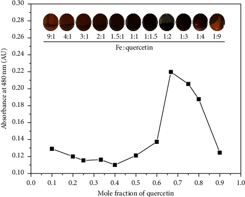
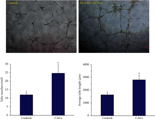
In cell therapy, contrast agents T1 and T2 are both needed for the labeling and tracking of transplanted stem cells over extended periods of time through magnetic resonance imaging (MRI). Importantly, the metal-quercetin complex via coordination chemistry has been studied extensively for biomedical applications, such as anticancer therapies and imaging probes. Herein, we report on the synthesis, characterization, and labeling of the iron (III)-quercetin complex, "IronQ," in circulating proangiogenic cells (CACs) and also explore tracking via the use of a clinical 1.5 Tesla (T) MRI scanner. Moreover, IronQ had a paramagnetic T1 positive contrast agent property with a saturation magnetization of 0.155 emu/g at 1.0 T and longitudinal relaxivity (r1) values of 2.29 and 3.70 mMs at 1.5 T for water and human plasma, respectively. Surprisingly, IronQ was able to promote CAC growth in conventional cell culture systems without the addition of specific growth factors. Increasing dosages of IronQ from 0 to 200 g/mL led to higher CAC uptake, and maximum labeling time was achieved in 10 days. The accumulated IronQ in CACs was measured by two methodologies, an inductively coupled plasma optical emission spectrometry (ICP-EOS) and T1-weighted MRI. In our research, we confirmed that IronQ has excellent dual functions with the use of an imaging probe for MRI. IronQ can also act as a stimulating agent by favoring circulating proangiogenic cell differentiation. Optimistically, IronQ is considered beneficial for alternative labeling and in the tracking of circulation proangiogenic cells and/or other stem cells in applications of cell therapy through noninvasive magnetic resonance imaging in both preclinical and clinical settings.
在细胞治疗中,对比剂 T1 和 T2 都需要用于通过磁共振成像 (MRI) 对移植的干细胞进行长时间的标记和跟踪。重要的是,通过配位化学,金属-槲皮素配合物已被广泛研究用于生物医学应用,如抗癌疗法和成像探针。在此,我们报告了铁 (III)-槲皮素配合物“铁 Q”的合成、表征和标记,以及在循环促血管生成细胞 (CAC) 中的追踪,并探索了使用临床 1.5 特斯拉 (T) MRI 扫描仪进行追踪。此外,IronQ 具有顺磁 T1 阳性对比剂特性,在 1.0 T 时饱和磁化强度为 0.155 emu/g,在 1.5 T 时水和人血浆中的纵向弛豫率 (r1) 值分别为 2.29 和 3.70 mMs。令人惊讶的是,IronQ 能够在没有添加特定生长因子的情况下促进 CAC 在常规细胞培养系统中的生长。IronQ 剂量从 0 增加到 200 μg/mL 会导致 CAC 摄取增加,并且在 10 天内达到最大标记时间。通过两种方法学,电感耦合等离子体发射光谱 (ICP-EOS) 和 T1 加权 MRI 测量了 CAC 中累积的 IronQ。在我们的研究中,我们证实了 IronQ 具有出色的双重功能,可用作 MRI 的成像探针。IronQ 还可以通过有利于循环促血管生成细胞分化来充当刺激剂。乐观地说,考虑到通过非侵入性磁共振成像在临床前和临床环境中进行细胞治疗的应用中,IronQ 有利于替代标记和循环促血管生成细胞和/或其他干细胞的追踪。