Sánchez Francisco J, Gonzalez Valeria A, Farrando Martin, Baigorria Jayat Ariel O, Segovia-Roldan Margarita, García-Mendívil Laura, Ordovás Laura, Prado Natalia J, Pueyo Esther, Diez Emiliano R
Department of Morphophysiology, School of Medicine, National University of Cuyo, Centro Universitario, Mendoza 5500, Argentina.
Department of Cardiovascular Surgery, Clinic of Cuyo, Mendoza 5500, Argentina.
Oxid Med Cell Longev. 2020 Dec 30;2020:8895078. doi: 10.1155/2020/8895078. eCollection 2020.
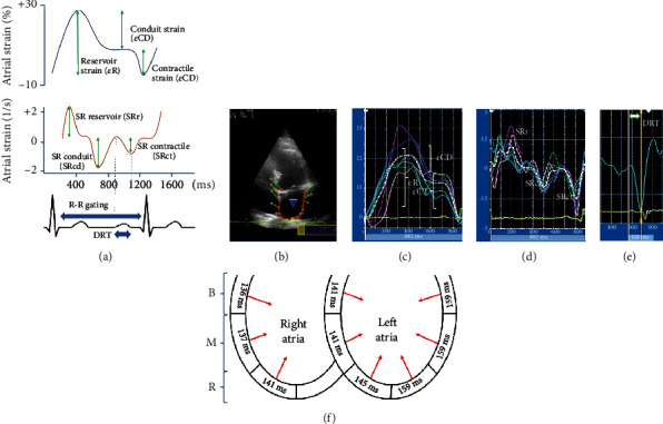
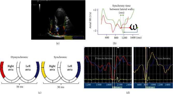
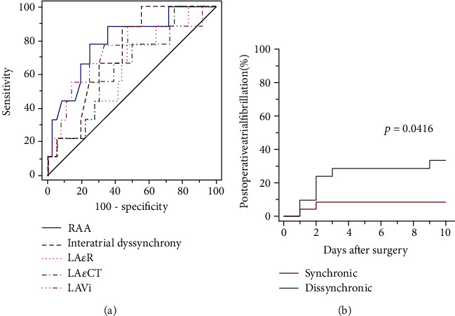
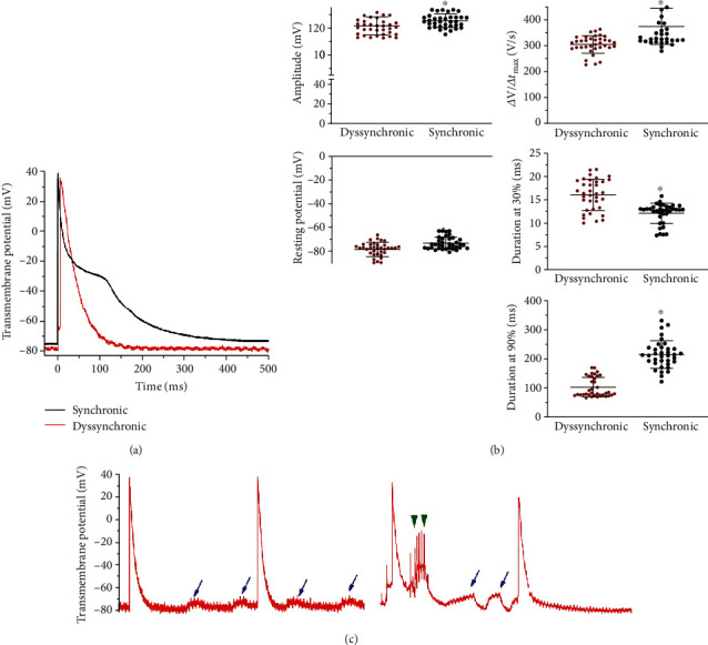
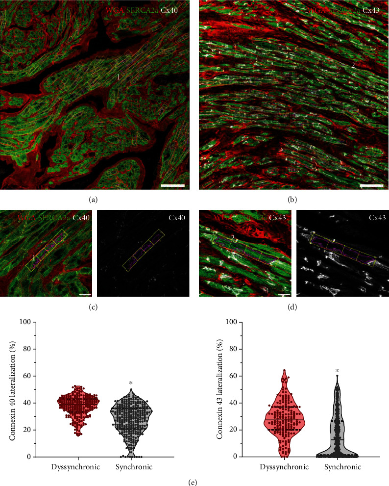
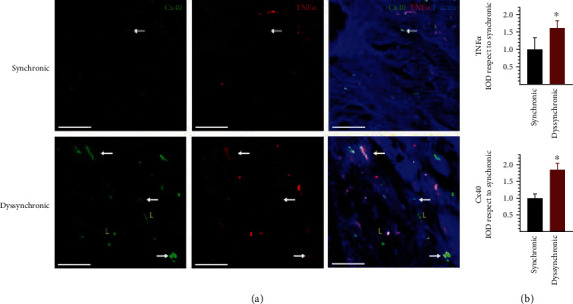
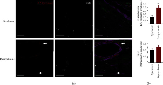
Aging leads to structural and electrophysiological changes that increase the risk of postoperative atrial arrhythmias; however, noninvasive preoperative markers of atrial proarrhythmic conditions are still needed. This study is aimed at assessing whether interatrial dyssynchrony determined using two-dimensional speckle tracking echocardiography relates to proarrhythmic structural and functional remodeling. A cohort of 45 patients in sinus rhythm referred for cardiac surgery was evaluated by echocardiography and surface electrocardiogram the day before the intervention. Transmembrane potential, connexin, and potassium channel distribution, inflammatory, and nitrooxidative markers were measured from right atrial tissue obtained from patients. A difference greater than 40 milliseconds between right and left atrial free wall contraction confirmed the presence of interatrial dyssynchrony in 21 patients. No difference in relation with age, previous diseases, and 2-dimensional echocardiographic findings as well as average values of global longitudinal right and left atrial strain were found between synchronic and dyssynchronic patients. Postoperative atrial fibrillation incidence increased from 8.3% in the synchronic group to 33.3% in the dyssynchronic ones. P wave duration showed no difference between groups. Action potentials from dyssynchronous patients decreased in amplitude, maximal rate of depolarization, and hyperpolarized. Duration at 30% of repolarization increased, being markedly shorter at 90% of repolarization. Only the dyssynchronous group showed early and delayed afterdepolarizations. Atrial tissue of dyssynchronous patients displayed lateralization of connexin 40 and increased connexin 43 expression and accumulation of tumor necrosis factor- in the intercalated disc. Tumor necrosis factor- did not colocalize, however, with lateralized connexin 40. Nitroxidative marks and K channels increased perivascularly and in myocytes. Our results demonstrate that, as compared to a traditional surface electrocardiogram, the novel noninvasive echocardiographic evaluation of interatrial dyssynchrony provides a better identification of nonaged-related proarrhythmic atrial remodeling with increased susceptibility to postoperative atrial fibrillation.
衰老会导致结构和电生理变化,增加术后房性心律失常的风险;然而,仍需要术前无创的房性心律失常状态标志物。本研究旨在评估使用二维斑点追踪超声心动图测定的心房不同步是否与心律失常的结构和功能重塑有关。对45例因心脏手术而窦性心律的患者在干预前一天进行超声心动图和体表心电图评估。从患者获取的右心房组织中测量跨膜电位、连接蛋白和钾通道分布、炎症和氧化亚氮标志物。右心房和左心房游离壁收缩之间的差异大于40毫秒证实21例患者存在心房不同步。在同步和不同步患者之间,未发现与年龄、既往疾病、二维超声心动图结果以及右心房和左心房整体纵向应变平均值相关的差异。术后房颤发生率从同步组的8.3%增加到不同步组的33.3%。两组间P波时限无差异。不同步患者的动作电位振幅、最大去极化速率降低且出现超极化。复极化30%时的时程增加,而复极化90%时明显缩短。只有不同步组显示早期和延迟后去极化。不同步患者的心房组织显示连接蛋白40侧向化,连接蛋白43表达增加,且肿瘤坏死因子在闰盘处积聚。然而,肿瘤坏死因子与侧向化的连接蛋白40不共定位。氧化亚氮标志物和钾通道在血管周围和心肌细胞中增加。我们的结果表明,与传统体表心电图相比,对心房不同步进行新型无创超声心动图评估能更好地识别与年龄无关的心律失常性心房重塑,且术后房颤易感性增加。