Kim Dae-Young, Moon Sun-Ha, Han Jang-Ho, Kim Mi-Jeong, Oh Seong-Ju, Bharti Dinesh, Lee Sung-Ho, Park Jong-Kuen, Rho Gyu-Jin, Jeon Byeong-Gyun
Department of Biology Education, Gyeongsang National University, Jinju, Republic of Korea.
OBS/Theriogenology and Biotechnology, Gyeongsang National University, Jinju, Republic of Korea.
Anim Cells Syst (Seoul). 2020 Dec 2;24(6):329-340. doi: 10.1080/19768354.2020.1847731.
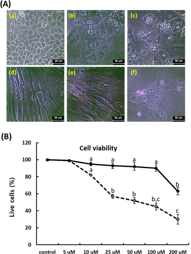
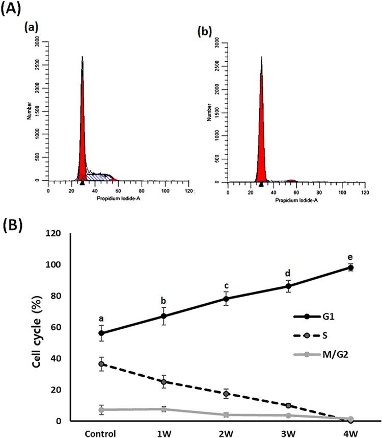
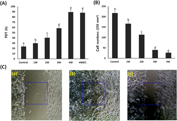
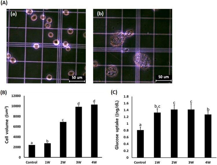
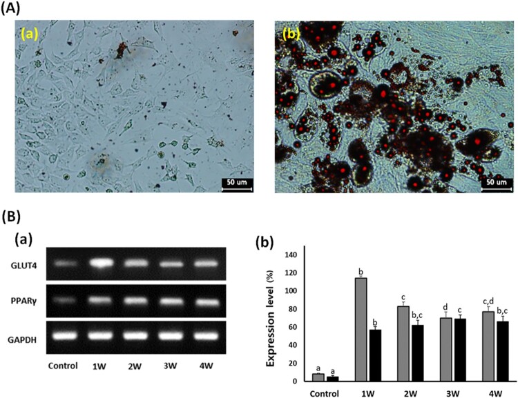
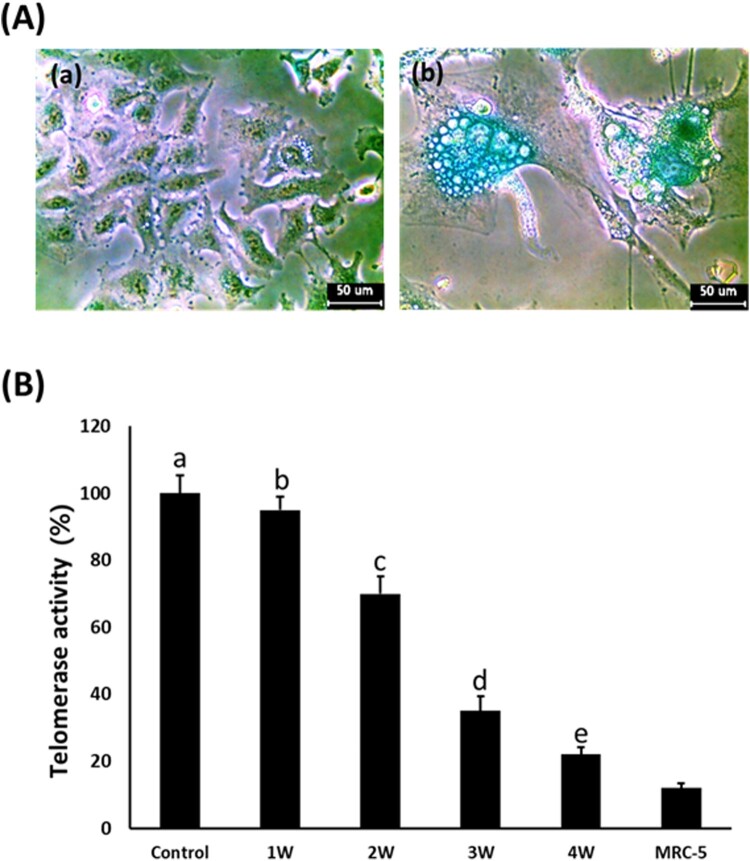
The present study investigated the terminal differentiation capacity into adipocytes and subsequent growth inhibition in A549 cancer cells treated with pioglitazone (PGZ), a PPARγ activator. The rate of cell growth in A549 cells was significantly (< .05) inhibited in concentrations above 10 μM PGZ while maintaining less cytotoxic effects in MRC-5 fibroblasts. Following 50 μM PGZ treatment, population doubling time (PDT) was significantly ( < .05) increased by inhibition of cell growth, as per increasing PGZ exposure time by up to 4 weeks. The adiposome-like vesicles were commonly observed in the PGZ-treated A549 cells, and the vesicles were highly stained with Oil-Red O solution. In addition, the cell size and expression of GLUT4 and PPARγ were significantly ( < .05) increased, as per increasing PGZ exposure time by up to 4 weeks. The significant ( < .05) down-regulation of telomerase activity and up-regulation of senescence-associated β-galactosidase (SA β-GAL) activity was displayed in the PGZ-treated A549 cells, as per increasing PGZ exposure time by up to 4 weeks. The G1 phase of the cell cycle was also significantly ( < .05) increased in the PGZ-treated A549 cells compared with untreated A549 cells. The present results have demonstrated that activation of PPARγ using PGZ induces cellular differentiation into adipocytes and inhibits cell growth in the A549 cancer cells. The terminal differentiation into adipocytes could offer potent chemotherapy in the cancer cells showing high glucose metabolism.
本研究调查了用吡格列酮(PGZ,一种PPARγ激活剂)处理的A549癌细胞向脂肪细胞的终末分化能力及随后的生长抑制情况。当PGZ浓度高于10 μM时,A549细胞的生长速率受到显著抑制(<0.05),而对MRC-5成纤维细胞的细胞毒性作用较小。在50 μM PGZ处理后,随着PGZ暴露时间延长至4周,细胞生长受到抑制,群体倍增时间(PDT)显著增加(<0.05)。在经PGZ处理的A549细胞中常见脂滴样小泡,这些小泡被油红O溶液高度染色。此外,随着PGZ暴露时间延长至4周,细胞大小以及GLUT4和PPARγ的表达显著增加(<0.05)。随着PGZ暴露时间延长至4周,经PGZ处理的A549细胞中端粒酶活性显著下调(<0.05),衰老相关β-半乳糖苷酶(SA β-GAL)活性上调。与未处理的A549细胞相比,经PGZ处理的A549细胞的细胞周期G1期也显著增加(<0.05)。目前的结果表明,使用PGZ激活PPARγ可诱导A549癌细胞向脂肪细胞分化并抑制其生长。向脂肪细胞的终末分化可为显示高糖代谢的癌细胞提供有效的化疗方法。