Grisaru-Tal Sharon, Itan Michal, Grass Daniel G, Torres-Roca Javier, Eschrich Steven A, Gordon Yaara, Dolitzky Avishay, Hazut Inbal, Avlas Shmuel, Jacobsen Elizabeth A, Ziv-Baran Tomer, Munitz Ariel
Department of Clinical Microbiology and Immunology, Sackler School of Medicine, Tel Aviv University, Tel-Aviv, Israel.
Department of Radiation Oncology, H. Lee Moffitt Cancer Center and Research Institute, Tampa, FL, USA.
Oncoimmunology. 2020 Dec 30;10(1):1859732. doi: 10.1080/2162402X.2020.1859732.
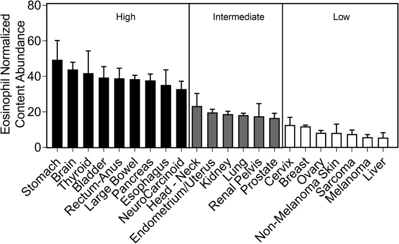
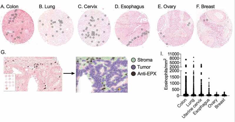
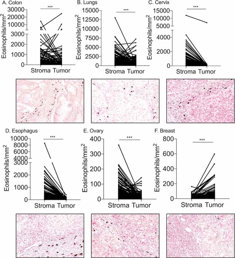
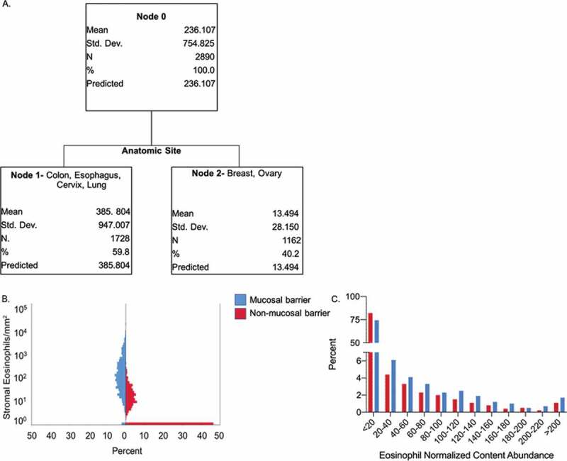
Eosinophils are bone marrow-derived granulocytes that display key effector functions in allergic diseases. Nonetheless, recent data highlight important roles for eosinophils in the tumor microenvironment (TME). Eosinophils have been attributed with pleiotropic and perhaps conflicting functions, which may be attributed at least in part to variations in eosinophil quantitation in the TME. Thus, a reliable, quantitative, and robust method for the assessment of eosinophilic infiltration in the TME is required. This type of methodology could standardize the identification of these cells and promote the subsequent generation of hypothesis-driven mechanistic studies. To this end, we conducted a comprehensive analysis of multiple primary tumors from distinct anatomical sites using a standardized method. Bioinformatics analysis of 10,469 genomically profiled primary tumors revealed that eosinophil abundance within different tumors can be categorized into three groups representing tumors with high, intermediate, and low eosinophil levels. Consequently, eosinophil abundance, as well as spatial distribution, was determined in tissue tumor arrays of six tumors representing all three classifications (colon and esophagus - high; lung - intermediate; cervix, ovary, and breast - low). With the exception of breast cancer, eosinophils were mainly localized in the tumor stroma. Importantly, the tumor anatomical site was identified as the primary predictive factor of eosinophil stromal density highlighting a distinction between mucosal-barrier organs versus non-mucosal barrier organs. These findings enhance our understanding of eosinophil diversity in the TME and provide a compelling rationale for future experiments assessing the activity of these cells.
嗜酸性粒细胞是源自骨髓的粒细胞,在过敏性疾病中发挥关键效应功能。尽管如此,最近的数据凸显了嗜酸性粒细胞在肿瘤微环境(TME)中的重要作用。嗜酸性粒细胞具有多效性且可能相互矛盾的功能,这至少部分可归因于TME中嗜酸性粒细胞定量的差异。因此,需要一种可靠、定量且稳健的方法来评估TME中的嗜酸性粒细胞浸润。这种方法可以使这些细胞的鉴定标准化,并促进后续基于假设的机制研究的开展。为此,我们使用一种标准化方法对来自不同解剖部位的多个原发性肿瘤进行了全面分析。对10469个进行了基因组分析的原发性肿瘤的生物信息学分析表明,不同肿瘤内的嗜酸性粒细胞丰度可分为三组,分别代表嗜酸性粒细胞水平高、中、低的肿瘤。因此,在代表所有三种分类的六种肿瘤(结肠和食管 - 高;肺 - 中;宫颈、卵巢和乳腺 - 低)的组织肿瘤阵列中确定了嗜酸性粒细胞丰度以及空间分布。除乳腺癌外,嗜酸性粒细胞主要定位于肿瘤基质中。重要的是,肿瘤解剖部位被确定为嗜酸性粒细胞基质密度的主要预测因素,突出了粘膜屏障器官与非粘膜屏障器官之间的区别。这些发现加深了我们对TME中嗜酸性粒细胞多样性的理解,并为未来评估这些细胞活性的实验提供了令人信服的理论依据。