• 文献检索
  • 文档翻译
  • 深度研究
  • 学术资讯
  • Suppr Zotero 插件Zotero 插件
  • 邀请有礼
  • 套餐&价格
  • 历史记录
应用&插件
Suppr Zotero 插件Zotero 插件浏览器插件Mac 客户端Windows 客户端微信小程序
定价
高级版会员购买积分包购买API积分包
服务
文献检索文档翻译深度研究API 文档MCP 服务
关于我们
关于 Suppr公司介绍联系我们用户协议隐私条款
关注我们

Suppr 超能文献

核心技术专利:CN118964589B侵权必究
粤ICP备2023148730 号-1Suppr @ 2026

文献检索

告别复杂PubMed语法,用中文像聊天一样搜索,搜遍4000万医学文献。AI智能推荐,让科研检索更轻松。

立即免费搜索

文件翻译

保留排版,准确专业,支持PDF/Word/PPT等文件格式,支持 12+语言互译。

免费翻译文档

深度研究

AI帮你快速写综述,25分钟生成高质量综述,智能提取关键信息,辅助科研写作。

立即免费体验

营养传感器 mTOR 和 OGT:胰腺细胞细胞器稳态的协调者。

Nutrient Sensor mTOR and OGT: Orchestrators of Organelle Homeostasis in Pancreatic -Cells.

机构信息

Department of Integrative Biology & Physiology, University of Minnesota Medical School, University of Minnesota, Minneapolis, Minnesota, USA.

Department of Surgery, University of Minnesota Medical School, University of Minnesota, Minneapolis, Minnesota, USA.

出版信息

J Diabetes Res. 2020 Dec 16;2020:8872639. doi: 10.1155/2020/8872639. eCollection 2020.

DOI:10.1155/2020/8872639
PMID:33457426
原文链接:https://pmc.ncbi.nlm.nih.gov/articles/PMC7787834/
Abstract

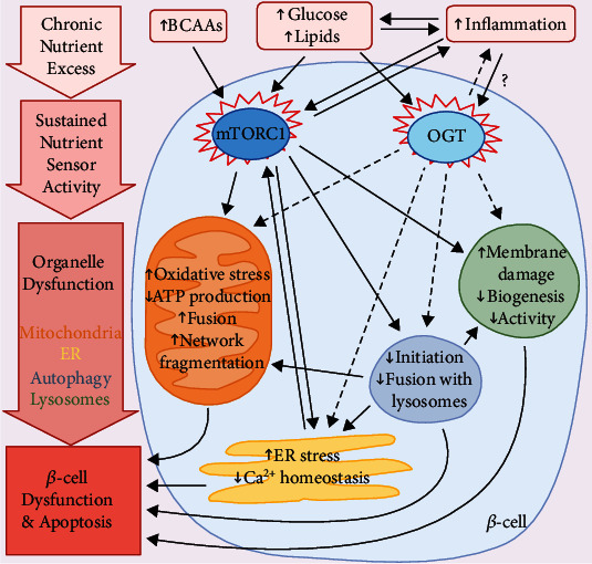
The purpose of this review is to integrate the role of nutrient-sensing pathways into -cell organelle dysfunction prompted by nutrient excess during type 2 diabetes (T2D). T2D encompasses chronic hyperglycemia, hyperlipidemia, and inflammation, which each contribute to -cell failure. These factors can disrupt the function of critical -cell organelles, namely, the ER, mitochondria, lysosomes, and autophagosomes. Dysfunctional organelles cause defects in insulin synthesis and secretion and activate apoptotic pathways if homeostasis is not restored. In this review, we will focus on mTORC1 and OGT, two major anabolic nutrient sensors with important roles in -cell physiology. Though acute stimulation of these sensors frequently improves -cell function and promotes adaptation to cell stress, chronic and sustained activity disturbs organelle homeostasis. mTORC1 and OGT regulate organelle function by influencing the expression and activities of key proteins, enzymes, and transcription factors, as well as by modulating autophagy to influence clearance of defective organelles. In addition, mTORC1 and OGT activity influence islet inflammation during T2D, which can further disrupt organelle and -cell function. Therapies for T2D that fine-tune the activity of these nutrient sensors have yet to be developed, but the important role of mTORC1 and OGT in organelle homeostasis makes them promising targets to improve -cell function and survival.

摘要

这篇综述的目的是整合营养感应途径在 2 型糖尿病(T2D)期间营养过剩引起的 -细胞细胞器功能障碍中的作用。T2D 包括慢性高血糖、高血脂和炎症,这些都促成了 -细胞衰竭。这些因素会破坏关键的 -细胞细胞器的功能,即内质网、线粒体、溶酶体和自噬体。如果不能恢复体内平衡,功能失调的细胞器会导致胰岛素合成和分泌缺陷,并激活凋亡途径。在这篇综述中,我们将重点讨论 mTORC1 和 OGT,这两种主要的合成代谢营养传感器在 -细胞生理学中具有重要作用。尽管这些传感器的急性刺激经常改善 -细胞功能并促进对细胞应激的适应,但慢性和持续的活动会扰乱细胞器的体内平衡。mTORC1 和 OGT 通过影响关键蛋白质、酶和转录因子的表达和活性,以及通过调节自噬来影响有缺陷的细胞器的清除,来调节细胞器功能。此外,mTORC1 和 OGT 的活性会影响 T2D 期间胰岛的炎症,这会进一步破坏细胞器和 -细胞功能。尚未开发出微调这些营养传感器活性的 T2D 疗法,但 mTORC1 和 OGT 在细胞器体内平衡中的重要作用使它们成为改善 -细胞功能和生存的有前途的靶点。

https://cdn.ncbi.nlm.nih.gov/pmc/blobs/bbc3/7787834/30d8a6f84a11/JDR2020-8872639.001.jpg

相似文献

1
Nutrient Sensor mTOR and OGT: Orchestrators of Organelle Homeostasis in Pancreatic -Cells.营养传感器 mTOR 和 OGT:胰腺细胞细胞器稳态的协调者。
J Diabetes Res. 2020 Dec 16;2020:8872639. doi: 10.1155/2020/8872639. eCollection 2020.
2
Role of the mammalian target of rapamycin (mTOR) complexes in pancreatic β-cell mass regulation.雷帕霉素哺乳动物靶点(mTOR)复合物在胰腺β细胞量调节中的作用。
Vitam Horm. 2014;95:425-69. doi: 10.1016/B978-0-12-800174-5.00017-X.
3
Proper mTORC1 Activity Is Required for Glucose Sensing and Early Adaptation in Human Pancreatic β Cells.适当的 mTORC1 活性是人类胰腺β细胞葡萄糖感应和早期适应所必需的。
J Clin Endocrinol Metab. 2021 Jan 23;106(2):e562-e572. doi: 10.1210/clinem/dgaa786.
4
Inhibition of PHLPP1/2 phosphatases rescues pancreatic β-cells in diabetes.抑制 PHLPP1/2 磷酸酶可挽救糖尿病中的胰岛β细胞。
Cell Rep. 2021 Aug 3;36(5):109490. doi: 10.1016/j.celrep.2021.109490.
5
mTOR controls ChREBP transcriptional activity and pancreatic β cell survival under diabetic stress.在糖尿病应激状态下,哺乳动物雷帕霉素靶蛋白(mTOR)控制碳水化合物反应元件结合蛋白(ChREBP)的转录活性及胰腺β细胞存活。
J Cell Biol. 2017 Jul 3;216(7):2091-2105. doi: 10.1083/jcb.201701085. Epub 2017 Jun 12.
6
Lysosomes in nutrient signalling: A focus on pancreatic β-cells.营养信号转导中的溶酶体:以胰腺β细胞为例。
Diabetes Obes Metab. 2018 Sep;20 Suppl 2:104-115. doi: 10.1111/dom.13389.
7
Loss of O-GlcNAcylation modulates mTORC1 and autophagy in β cells, driving diabetes 2 progression.O-连接的N-乙酰葡糖胺糖基化修饰的缺失会调节β细胞中的mTORC1和自噬,推动2型糖尿病的进展。
JCI Insight. 2024 Dec 6;9(23):e183033. doi: 10.1172/jci.insight.183033.
8
Bridges between mitochondrial oxidative stress, ER stress and mTOR signaling in pancreatic β cells.胰腺β细胞中线粒体氧化应激、内质网应激与mTOR信号通路之间的桥梁。
Cell Signal. 2016 Aug;28(8):1099-104. doi: 10.1016/j.cellsig.2016.05.007. Epub 2016 May 14.
9
Inhibition of autophagic turnover in β-cells by fatty acids and glucose leads to apoptotic cell death.脂肪酸和葡萄糖对β细胞自噬周转的抑制会导致细胞凋亡死亡。
J Biol Chem. 2015 Mar 6;290(10):6071-85. doi: 10.1074/jbc.M114.605345. Epub 2014 Dec 29.
10
Loss of mTORC1 signalling impairs β-cell homeostasis and insulin processing.mTORC1 信号的丧失会损害β细胞的内稳态和胰岛素的加工。
Nat Commun. 2017 Jul 12;8:16014. doi: 10.1038/ncomms16014.

引用本文的文献

1
Sex Differences in Pancreatic β-Cell Physiology and Glucose Homeostasis in C57BL/6J Mice.C57BL/6J小鼠胰腺β细胞生理学和葡萄糖稳态中的性别差异
J Endocr Soc. 2023 Jul 25;7(9):bvad099. doi: 10.1210/jendso/bvad099. eCollection 2023 Aug 2.
2
β Cell and Autophagy: What Do We Know?β 细胞与自噬:我们了解多少?
Biomolecules. 2023 Apr 4;13(4):649. doi: 10.3390/biom13040649.
3
Yo-Yo Dieting: Mixed Messages for β-Cell Plasticity.溜溜球节食法:对β细胞可塑性的复杂信号

本文引用的文献

1
AMPK Profiling in Rodent and Human Pancreatic Beta-Cells under Nutrient-Rich Metabolic Stress.营养丰富的代谢应激下啮齿动物和人胰岛β细胞中 AMPK 的分析。
Int J Mol Sci. 2020 Jun 1;21(11):3982. doi: 10.3390/ijms21113982.
2
Potential positive and negative consequences of ZnT8 inhibition.ZnT8 抑制的潜在正反两方面影响。
J Endocrinol. 2020 Aug;246(2):189-205. doi: 10.1530/JOE-20-0138.
3
Islet O-GlcNAcylation Is Required for Lipid Potentiation of Insulin Secretion through SERCA2.胰岛 O-GlcNAc 修饰对于通过 SERCA2 增强胰岛素分泌的脂质作用是必需的。
Diabetes. 2022 Nov 1;71(11):2253-2255. doi: 10.2337/dbi22-0024.
4
Metabolomic Profiling of Amino Acids in Human Plasma Distinguishes Diabetic Kidney Disease From Type 2 Diabetes Mellitus.人血浆中氨基酸的代谢组学分析可区分糖尿病肾病与2型糖尿病。
Front Med (Lausanne). 2021 Nov 29;8:765873. doi: 10.3389/fmed.2021.765873. eCollection 2021.
5
Identification of Novel Regulatory Regions Induced by Intrauterine Growth Restriction in Rat Islets.鉴定宫内生长受限诱导的大鼠胰岛新型调控区域。
Endocrinology. 2022 Feb 1;163(2). doi: 10.1210/endocr/bqab251.
Cell Rep. 2020 May 5;31(5):107609. doi: 10.1016/j.celrep.2020.107609.
4
A Nutrient-Sensing Transition at Birth Triggers Glucose-Responsive Insulin Secretion.出生时的营养感应转变触发葡萄糖响应性胰岛素分泌。
Cell Metab. 2020 May 5;31(5):1004-1016.e5. doi: 10.1016/j.cmet.2020.04.004.
5
Mitochondrial Mechanisms in Diabetic Cardiomyopathy.糖尿病心肌病中的线粒体机制。
Diabetes Metab J. 2020 Feb;44(1):33-53. doi: 10.4093/dmj.2019.0185.
6
Nutrient-Induced Metabolic Stress, Adaptation, Detoxification, and Toxicity in the Pancreatic β-Cell.营养诱导的胰腺β细胞代谢应激、适应、解毒和毒性。
Diabetes. 2020 Mar;69(3):279-290. doi: 10.2337/dbi19-0014.
7
Modulation of O-GlcNAcylation Regulates Autophagy in Cortical Astrocytes.O-GlcNAcylation 修饰调节皮质星形胶质细胞中的自噬。
Oxid Med Cell Longev. 2019 Nov 13;2019:6279313. doi: 10.1155/2019/6279313. eCollection 2019.
8
Obesity-Induced Regulator of Calcineurin 1 Overexpression Leads to β-Cell Failure Through Mitophagy Pathway Inhibition.肥胖诱导钙调神经磷酸酶 1 过表达通过抑制线粒体自噬途径导致β细胞衰竭。
Antioxid Redox Signal. 2020 Mar 1;32(7):413-428. doi: 10.1089/ars.2019.7806.
9
Rapamycin Attenuates High Glucose-Induced Inflammation Through Modulation of mTOR/NF-κB Pathways in Macrophages.雷帕霉素通过调节巨噬细胞中的mTOR/NF-κB信号通路减轻高糖诱导的炎症。
Front Pharmacol. 2019 Oct 30;10:1292. doi: 10.3389/fphar.2019.01292. eCollection 2019.
10
The relevance of AMP-activated protein kinase in insulin-secreting β cells: a potential target for improving β cell function?AMP 激活的蛋白激酶在胰岛素分泌β细胞中的相关性:改善β细胞功能的潜在靶点?
J Physiol Biochem. 2019 Nov;75(4):423-432. doi: 10.1007/s13105-019-00706-3. Epub 2019 Nov 5.