Kemppainen Reko, Suilamo Sami, Ranta Iiro, Pesola Marko, Halkola Aleksi, Eufemio Alvin, Minn Heikki, Keyriläinen Jani
Philips Oy, Äyritie 4, FI-01510 Vantaa, Finland.
Department of Neuroscience and Biomedical Engineering, Aalto University School of Science, Rakentajanaukio 2 C, FI-02150 Espoo, Finland.
Phys Imaging Radiat Oncol. 2019 Jun 22;11:1-8. doi: 10.1016/j.phro.2019.06.001. eCollection 2019 Jul.
The clinical feasibility of synthetic computed tomography (sCT) images derived from magnetic resonance imaging (MRI) images for external beam radiation therapy (EBRT) planning have been studied and adopted into clinical use recently. This paper evaluates the dosimetric and positioning performance of a sCT approach for different pelvic cancers.
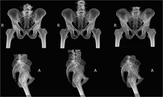
Seventy-five patients receiving EBRT at Turku University Hospital (Turku, Finland) were enrolled in the study. The sCT images were generated as part of a clinical MRI-simulation procedure. Dose calculation accuracy was assessed by comparing the sCT-based calculation with a CT-based calculation. In addition, we evaluated the patient position verification accuracy for both digitally reconstructed radiograph (DRR) and cone beam computed tomography (CBCT) -based image guidance using a subset of the cohort. Furthermore, the relevance of using continuous Hounsfield unit values was assessed.
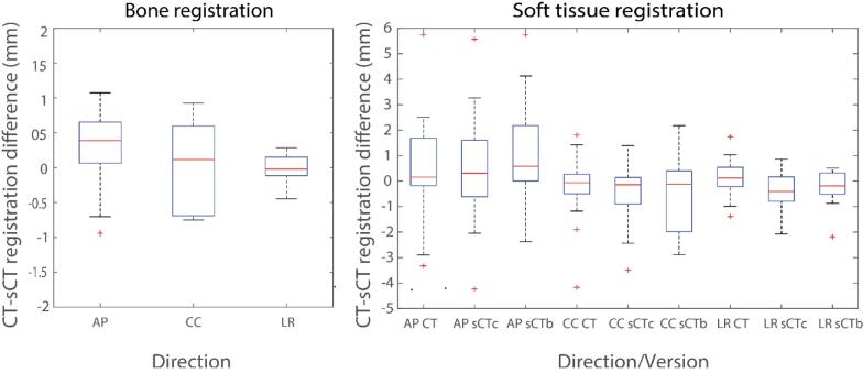
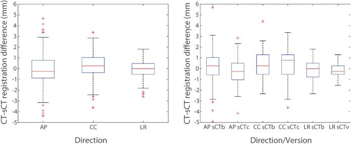
The mean (standard deviation) relative dose difference in the planning target volume mean dose computed over various cancer groups was less than 0.2 (0.4)% between sCT and CT. Among all groups, the average minimum gamma-index pass-rates were better than 95% with a 2%/2mm gamma-criteria. The difference between sCT- and CT-DRR-based patient positioning was less than 0.3 (1.4) mm in all directions. The registrations of sCT to CBCT produced similar results as compared with CT to CBCT registrations.
The use of sCT for clinical EBRT dose calculation and patient positioning in the investigated types of pelvic cancers was dosimetrically and geometrically accurate for clinical use.
源自磁共振成像(MRI)图像的合成计算机断层扫描(sCT)图像用于外照射放疗(EBRT)计划的临床可行性已得到研究,并于近期投入临床应用。本文评估了sCT方法对不同盆腔癌的剂量学和定位性能。
招募了75名在芬兰图尔库大学医院接受EBRT治疗的患者。sCT图像是作为临床MRI模拟程序的一部分生成的。通过将基于sCT的计算与基于CT的计算进行比较来评估剂量计算准确性。此外,我们使用队列中的一个子集评估了基于数字重建射线照相(DRR)和基于锥形束计算机断层扫描(CBCT)的图像引导的患者位置验证准确性。此外,还评估了使用连续Hounsfield单位值的相关性。
在不同癌症组中计算的计划靶体积平均剂量的平均(标准差)相对剂量差异在sCT和CT之间小于0.2(0.4)%。在所有组中,采用2%/2mm的伽马标准时,平均最小伽马指数通过率优于95%。基于sCT和CT-DRR的患者定位在各个方向上的差异均小于0.3(1.4)mm。与CT到CBCT的配准相比,sCT到CBCT的配准产生了相似的结果。
在研究的盆腔癌类型中,将sCT用于临床EBRT剂量计算和患者定位在剂量学和几何学上对于临床应用是准确的。