Fang Raymond, Mazur Thomas, Mutic Sasa, Khan Rao
Department of Radiation Oncology, Washington University School of Medicine, St. Louis, MO, USA.
Phys Imaging Radiat Oncol. 2018 Nov 2;8:1-7. doi: 10.1016/j.phro.2018.10.002. eCollection 2018 Oct.
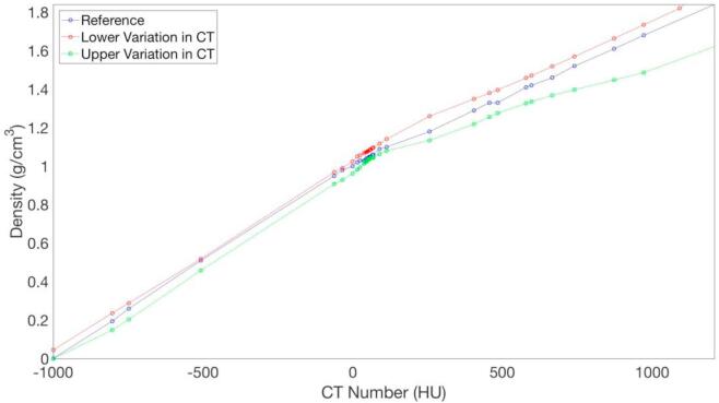
A key step in electron Monte Carlo dose calculation requires converting Computed Tomography (CT) numbers from a tomographic acquisition to a mass density. This study investigates the dosimetric consequences of perturbations applied to a calibration table between CT number and mass density.
A literature search was performed to define lower and upper bounds for physically reasonable perturbations to a reference CT number to mass density calibration table. Electron beam dose was calculated for ten patients using these variations and the results were compared to clinical plans originally derived with a reference calibration table. Dose differences both globally and in the Planning Target Volume (PTV) were assessed using dose- and volume-based metrics and 3- dimensional gamma analysis for each patient.
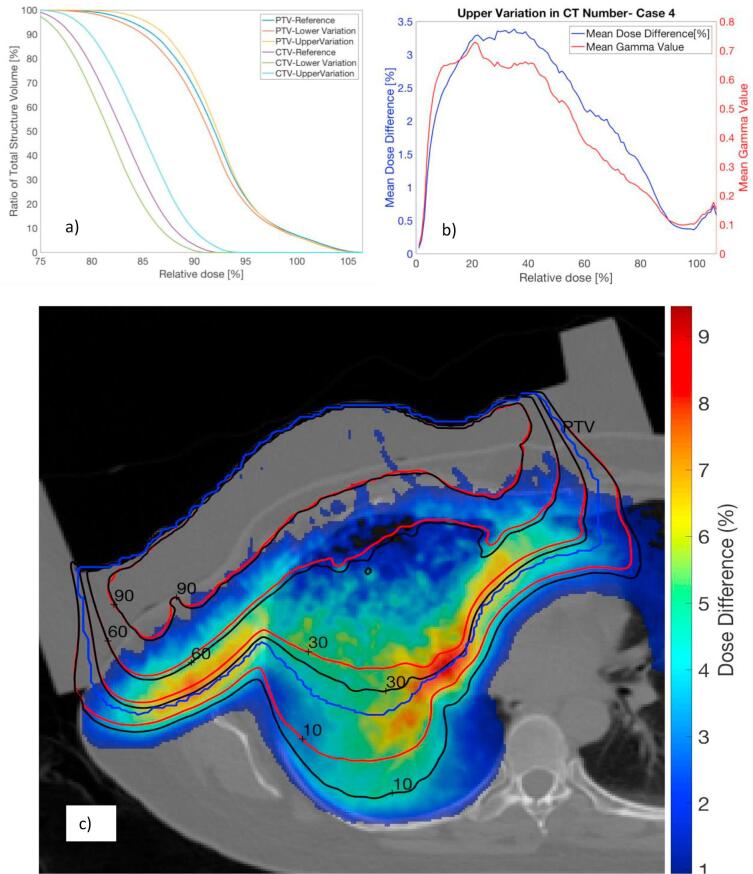
Small but statistically significant differences were observed between perturbations and reference data for certain metrics including volume of the 50% prescription isodose. Upper and lower variations in CT number to mass density calibration yielded mean values of V50% that were 4.4% larger and 2.1% smaller than reference values respectively. Gamma analysis using 3%/3mm criteria indicated >99% passing rate for the PTV for all patients. Global gamma analysis for some patients showed larger discrepancies possibly due to large electron path lengths through inhomogeneities.
In most patients, physically reasonable perturbations in CT number to mass density curves will not induce clinically significant impact on calculated target dose distributions. Strong dependence of electron transport on voxel material may produce dose speckle throughout the volume. Care should be taken in evaluating critical structures at depths beyond the target volume in highly heterogeneous regions.
电子蒙特卡罗剂量计算中的关键步骤是将断层扫描采集得到的计算机断层扫描(CT)数值转换为质量密度。本研究调查了对CT数值与质量密度校准表施加扰动所产生的剂量学后果。
进行文献检索以确定对参考CT数值至质量密度校准表进行物理上合理扰动的下限和上限。使用这些变化为10名患者计算电子束剂量,并将结果与最初使用参考校准表得出的临床计划进行比较。使用基于剂量和体积的指标以及对每位患者进行三维伽马分析,评估全局和计划靶区(PTV)中的剂量差异。
在某些指标上,包括50%处方等剂量线的体积,扰动与参考数据之间观察到了虽小但具有统计学意义的差异。CT数值至质量密度校准的上下变化分别产生了比参考值大4.4%和小2.1%的V50%平均值。使用3%/3mm标准的伽马分析表明,所有患者PTV的通过率均>99%。对一些患者的全局伽马分析显示差异较大,这可能是由于电子穿过不均匀性区域的路径长度较长所致。
在大多数患者中,CT数值至质量密度曲线的物理上合理的扰动不会对计算出的靶区剂量分布产生临床显著影响。电子传输对体素材料的强烈依赖性可能会在整个体积内产生剂量斑点。在评估高度异质区域中靶区体积深度以外的关键结构时应谨慎。