Department of Orthopaedics, Xinqiao Hospital, Army Medical University (Third Military Medical University), Chongqing, People's Republic of China.
Institute of Immunology, PLA, Army Medical University (Third Military Medical University), Chongqing, People's Republic of China.
Stem Cells. 2021 Apr;39(4):467-481. doi: 10.1002/stem.3322. Epub 2021 Jan 18.
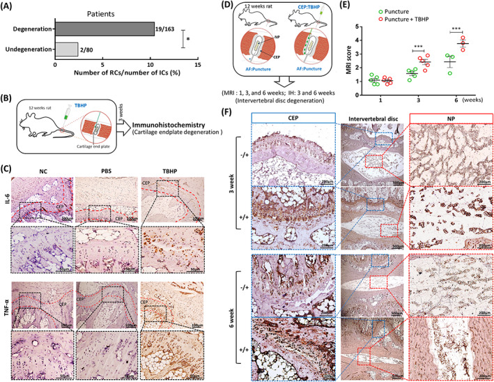
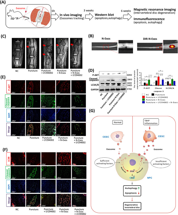
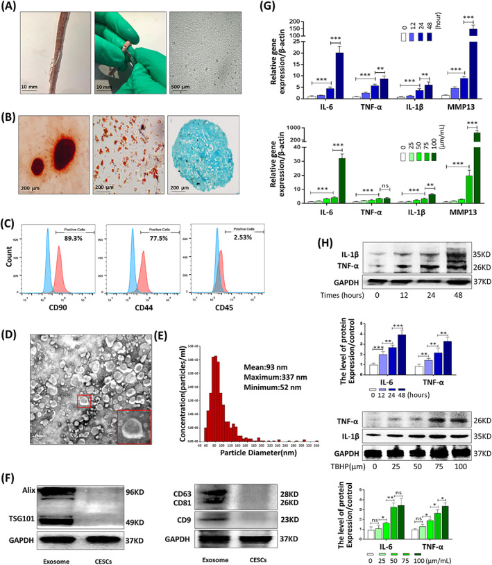
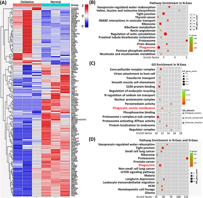
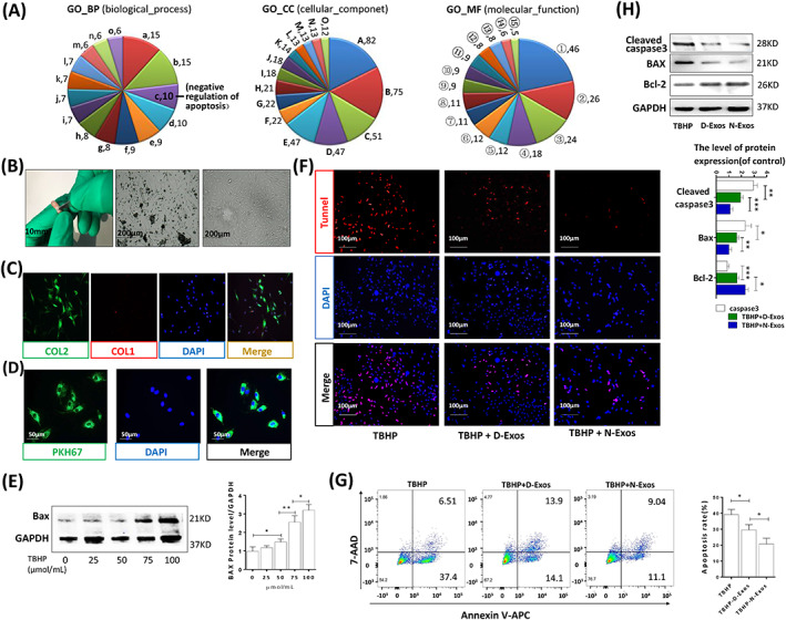
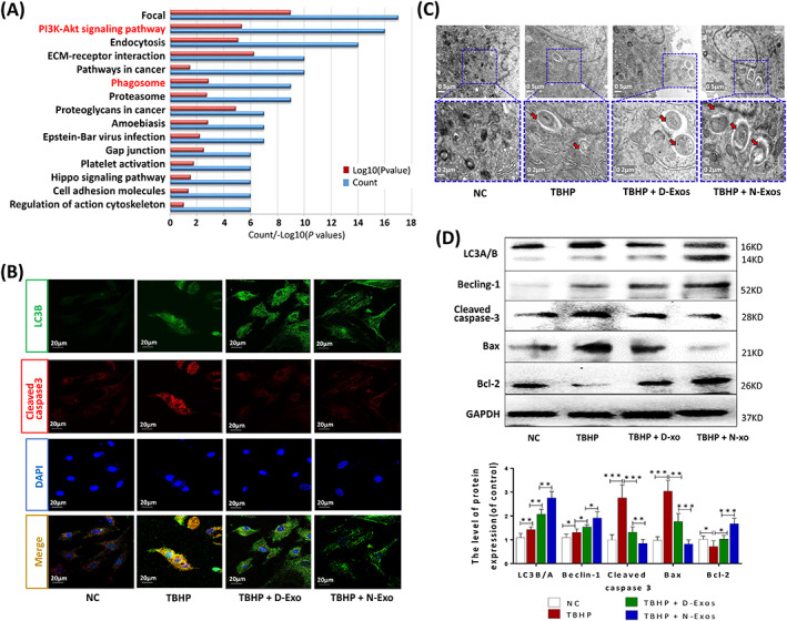
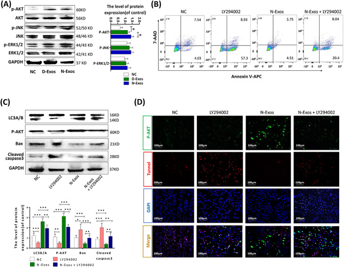
Degeneration of the cartilage endplate (CEP) induces intervertebral disc degeneration (IVDD). Nucleus pulposus cell (NPC) apoptosis is also an important exacerbating factor in IVDD, but the cascade mechanism in IVDD is not clear. We investigated the apoptosis of NPCs and IVDD when stimulated by normal cartilage endplate stem cell (CESC)-derived exosomes (N-Exos) and degenerated CESC-derived exosomes (D-Exos) in vitro and in vivo. Tert-butyl hydroperoxide (TBHP) was used to induce inflammation of CESCs. The bioinformatics differences between N-Exos and D-Exos were analyzed using mass spectrometry, heat map, and Kyoto Encyclopedia of Genes and Genomes (KEGG) enrichment analysis. NPC apoptosis was examined using TUNEL staining. The involvement of the AKT and autophagy signaling pathways was investigated using the signaling inhibitor LY294002. Magnetic resonance imaging, Western blotting, and immunofluorescence staining were used to evaluate the therapeutic effects of N-Exos in rats with IVDD. TBHP effectively induced inflammation and the degeneration of CEP in rat. N-Exos were more conducive to autophagy activation than D-Exos. The apoptotic rate of NPCs decreased obviously after treatment with N-Exos compared to D-Exos. N-Exos inhibited NPCs apoptosis and attenuated IVDD in rat via activation of the AKT and autophagy pathways. These results are the first findings to confirm that CEP delayed the progression of IVDD via exosomes. The therapeutic effects of N-Exos on NPC apoptosis inhibition and the slowing of IVDD progression were more effective than D-Exos due to activation of the PI3K/AKT/autophagy pathway, which explained the increase in the incidence of IVDD after inflammation of the CEP.
软骨终板(CEP)退变导致椎间盘退变(IVDD)。核 细胞(NPC)凋亡也是 IVDD 的一个重要加重因素,但 IVDD 的级联机制尚不清楚。我们研究了 NPC 在体外和体内受正常软骨终板干细胞(CESC)衍生外泌体(N-Exos)和退变 CESC 衍生外泌体(D-Exos)刺激时的凋亡以及 IVDD。叔丁基过氧化物(TBHP)用于诱导 CESCs 炎症。使用质谱、热图和京都基因与基因组百科全书(KEGG)富集分析分析 N-Exos 和 D-Exos 之间的生物信息学差异。使用 TUNEL 染色检查 NPC 凋亡。使用信号抑制剂 LY294002 研究 AKT 和自噬信号通路的参与情况。使用磁共振成像、Western blot 和免疫荧光染色评估 N-Exos 在 IVDD 大鼠中的治疗效果。TBHP 有效地诱导了大鼠 CEP 炎症和退变。N-Exos 比 D-Exos 更有利于自噬激活。与 D-Exos 相比,N-Exos 处理后 NPC 凋亡率明显降低。N-Exos 通过激活 AKT 和自噬通路抑制 NPC 凋亡并减轻大鼠 IVDD。这些结果是首次发现 CEP 通过外泌体延缓 IVDD 进展。由于激活了 PI3K/AKT/自噬通路,N-Exos 对 NPC 凋亡抑制和减缓 IVDD 进展的治疗效果比 D-Exos 更有效,这解释了 CEP 炎症后 IVDD 发生率增加的原因。