• 文献检索
  • 文档翻译
  • 深度研究
  • 学术资讯
  • Suppr Zotero 插件Zotero 插件
  • 邀请有礼
  • 套餐&价格
  • 历史记录
应用&插件
Suppr Zotero 插件Zotero 插件浏览器插件Mac 客户端Windows 客户端微信小程序
定价
高级版会员购买积分包购买API积分包
服务
文献检索文档翻译深度研究API 文档MCP 服务
关于我们
关于 Suppr公司介绍联系我们用户协议隐私条款
关注我们

Suppr 超能文献

核心技术专利:CN118964589B侵权必究
粤ICP备2023148730 号-1Suppr @ 2026

文献检索

告别复杂PubMed语法,用中文像聊天一样搜索,搜遍4000万医学文献。AI智能推荐,让科研检索更轻松。

立即免费搜索

文件翻译

保留排版,准确专业,支持PDF/Word/PPT等文件格式,支持 12+语言互译。

免费翻译文档

深度研究

AI帮你快速写综述,25分钟生成高质量综述,智能提取关键信息,辅助科研写作。

立即免费体验

UBM 引导下巩膜扣带术治疗伴有睫状体非色素上皮撕裂的 Schwartz-Matsuo 综合征:病例报告。

UBM-guided scleral buckling for Schwartz-Matsuo syndrome with tear of nonpigmented epithelium of the ciliary body: a case report.

机构信息

Eye Center, Second Affiliated Hospital, School of Medicine, Zhejiang University, No. 88 Jiefang Rd, Hangzhou, 310009, China.

Department of Ophthalmology, Zhuji People's Hospital of Zhejiang Province, 311800, Zhuji, China.

出版信息

BMC Ophthalmol. 2021 Jan 19;21(1):49. doi: 10.1186/s12886-021-01809-6.

DOI:10.1186/s12886-021-01809-6
PMID:33468097
原文链接:https://pmc.ncbi.nlm.nih.gov/articles/PMC7816455/
Abstract

BACKGROUND

Tears in Schwartz-Matsuo syndrome are generally confirmed by preoperative ophthalmoscopic examination. A case of Schwartz-Matsuo syndrome with a tear detected by ultrasound biomicroscopy (UBM) and treated by UBM-guided scleral buckling was reported, and its mechanism was analysed.

CASE PRESENTATION

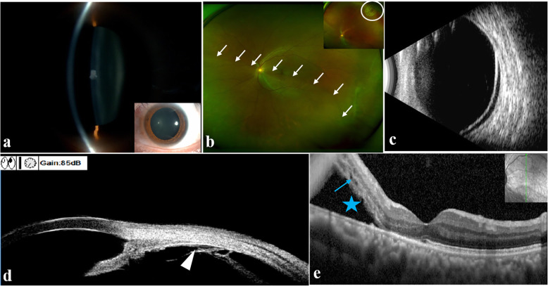
A 40-year-old Chinese man presented with blurry vision and intermittent eye pain in his left eye for three days. The visual acuity of the left eye decreased from 20/20 to 20/40, and the intraocular pressure (IOP) fluctuated dramatically from 24.0 mmHg to 56.7 mmHg at the first visit. Gonioscopy revealed that the chamber angle remained open. A macula-involving inferior retinal detachment extending from 4:30 to 9:30 with no obvious causative break was observed through ophthalmoscopic examination. However, a single small tear was detected at the nonpigmented epithelium of pars plana of the ciliary body at approximately 7-8 o'clock by UBM. The loss of photoreceptor outer segments and ellipsoid zone and the existence of macular microcysts in the inner and outer nuclear layers were observed in the detached macula by optical coherence tomography. Then, he underwent successful scleral buckling guided by UBM. Three months later, the retina was flat with normal IOP, and the best corrected visual acuity of his left eye gradually improved to 20/25. UBM confirmed the closure of the tear.

CONCLUSIONS

Tear of the nonpigmented epithelium of the ciliary body is a rare condition associated with Schwartz-Matsuo syndrome. UBM plays a key role in detecting occult tears of the nonpigmented epithelium of the ciliary body, guiding scleral buckling surgery, and observing the closure of the tear postoperatively.

摘要

背景

Schwartz-Matsuo 综合征的裂孔通常通过术前眼底检查来确诊。本文报告了一例超声生物显微镜(UBM)发现裂孔并经 UBM 引导巩膜扣带术治疗的 Schwartz-Matsuo 综合征病例,并分析其发病机制。

病例介绍

一名 40 岁的中国男性,因左眼视物模糊伴间歇性眼痛 3 天就诊。左眼视力从 20/20 下降至 20/40,眼压在首次就诊时从 24.0mmHg 波动至 56.7mmHg。房角镜检查发现房角开放。眼底镜检查发现黄斑下累及视网膜脱离,从 4:30 延伸至 9:30,未见明显裂孔。然而,UBM 检查发现睫状体平坦部无色素上皮处有单个小裂孔,约位于 7-8 点钟。光学相干断层扫描发现脱离的黄斑内层和外层核层存在光感受器外节和椭圆体带丢失以及黄斑微囊。随后,他接受了 UBM 引导的巩膜扣带术。3 个月后,视网膜平复,眼压正常,左眼最佳矫正视力逐渐提高至 20/25。UBM 证实裂孔已闭合。

结论

睫状体无色素上皮的裂孔是一种罕见的 Schwartz-Matsuo 综合征相关病变。UBM 在检测睫状体无色素上皮隐匿性裂孔、引导巩膜扣带术以及观察术后裂孔闭合方面发挥着关键作用。

https://cdn.ncbi.nlm.nih.gov/pmc/blobs/2a08/7816455/a6b0cfd3f9e6/12886_2021_1809_Fig3_HTML.jpg
https://cdn.ncbi.nlm.nih.gov/pmc/blobs/2a08/7816455/07e0a73b8af5/12886_2021_1809_Fig1_HTML.jpg
https://cdn.ncbi.nlm.nih.gov/pmc/blobs/2a08/7816455/0bca38af1f24/12886_2021_1809_Fig2_HTML.jpg
https://cdn.ncbi.nlm.nih.gov/pmc/blobs/2a08/7816455/a6b0cfd3f9e6/12886_2021_1809_Fig3_HTML.jpg
https://cdn.ncbi.nlm.nih.gov/pmc/blobs/2a08/7816455/07e0a73b8af5/12886_2021_1809_Fig1_HTML.jpg
https://cdn.ncbi.nlm.nih.gov/pmc/blobs/2a08/7816455/0bca38af1f24/12886_2021_1809_Fig2_HTML.jpg
https://cdn.ncbi.nlm.nih.gov/pmc/blobs/2a08/7816455/a6b0cfd3f9e6/12886_2021_1809_Fig3_HTML.jpg

相似文献

1
UBM-guided scleral buckling for Schwartz-Matsuo syndrome with tear of nonpigmented epithelium of the ciliary body: a case report.UBM 引导下巩膜扣带术治疗伴有睫状体非色素上皮撕裂的 Schwartz-Matsuo 综合征:病例报告。
BMC Ophthalmol. 2021 Jan 19;21(1):49. doi: 10.1186/s12886-021-01809-6.
2
Schwartz-Matsuo syndrome in retinal detachment with tears of the nonpigmented epithelium of the ciliary body.伴有睫状体无色素上皮撕裂的视网膜脱离中的施瓦茨-松尾综合征。
Acta Ophthalmol Scand. 1998 Aug;76(4):481-5. doi: 10.1034/j.1600-0420.1998.760417.x.
3
[Ciliochoroidal detachment following scleral buckling surgery for rhegmatogenous retinal detachment].[巩膜扣带术治疗孔源性视网膜脱离后睫状脉络膜脱离]
Nippon Ganka Gakkai Zasshi. 2000 May;104(5):344-8.
4
Ciliary body edema after scleral buckling surgery for rhegmatogenous retinal detachment.孔源性视网膜脱离巩膜扣带术后睫状体水肿
Ophthalmology. 2006 Jan;113(1):36-41. doi: 10.1016/j.ophtha.2005.09.023. Epub 2005 Nov 10.
5
Schwartz-Matsuo Syndrome施瓦茨-松尾综合征
6
Outcomes of scleral buckling using chandelier endoillumination.使用吊灯式眼内照明进行巩膜扣带术的结果
Acta Ophthalmol. 2017 Sep;95(6):591-594. doi: 10.1111/aos.13326. Epub 2016 Dec 14.
7
[A study on ocular anterior segment structure after scleral buckling surgery for retinal detachment].[视网膜脱离巩膜扣带术后眼前节结构的研究]
Zhonghua Yan Ke Za Zhi. 1999 Jul;35(4):309-11, 17.
8
Retinal detachments due to traumatic tears in the pars plana ciliaris.由于睫状体扁平部外伤性撕裂导致的视网膜脱离。
Retina. 1998;18(6):506-9. doi: 10.1097/00006982-199811000-00002.
9
Long ciliary processes with no ciliary sulcus and appositional angle closure assessed by ultrasound biomicroscopy.通过超声生物显微镜检查评估,发现睫状突长,无睫状沟且存在房角关闭。
J Glaucoma. 2006 Oct;15(5):371-9. doi: 10.1097/01.ijg.0000212251.72207.19.
10
Ciliochoroidal Detachment Following Scleral Buckling Surgery for Rhegmatogenous Retinal Detachment.孔源性视网膜脱离巩膜扣带术后的睫状体脉络膜脱离
Jpn J Ophthalmol. 2000 Nov 1;44(6):692-693. doi: 10.1016/s0021-5155(00)00255-0.

引用本文的文献

1
Ultrasound biomicroscopy-based diagnosis and cryotherapy for idiopathic rhegmatogenous retinal detachment secondary to pars plicata non-pigmented epithelium break: a case description.基于超声生物显微镜的原发性锯齿缘非色素上皮裂孔继发孔源性视网膜脱离的诊断与冷冻治疗:病例描述
Quant Imaging Med Surg. 2025 Aug 1;15(8):7618-7623. doi: 10.21037/qims-2025-329. Epub 2025 Jul 25.
2
Case report: Ultrasound biomicroscopy as a guide for the selection of injection sites for dexamethasone intravitreal implant (Ozurdex) for peripheral granulomatous ocular toxocariasis in children.病例报告:超声生物显微镜引导下为儿童周边肉芽肿性眼弓蛔虫病患者选择地塞米松玻璃体内植入剂(Ozurdex)的注射部位
Front Med (Lausanne). 2023 May 16;10:1176585. doi: 10.3389/fmed.2023.1176585. eCollection 2023.

本文引用的文献

1
Schwartz-Matsuo syndrome: An important cause of secondary glaucoma.施瓦茨-松尾综合征:继发性青光眼的一个重要病因。
Am J Ophthalmol Case Rep. 2020 Jan 3;17:100586. doi: 10.1016/j.ajoc.2020.100586. eCollection 2020 Mar.
2
Ultrasound biomicroscopy as a vital tool in occult phacomorphic glaucoma.超声生物显微镜作为隐匿性晶状体溶解性青光眼的重要工具。
Rom J Ophthalmol. 2019 Oct-Dec;63(4):311-314.
3
Diagnosis of Peripheral Retinoschisis Using Ultrasound Biomicroscopy.使用超声生物显微镜诊断周边视网膜劈裂症
Ophthalmic Surg Lasers Imaging Retina. 2019 Aug 1;50(8):e196-e202. doi: 10.3928/23258160-20190806-12.
4
Electron Microscopic Evidence of Photoreceptor Outer-segments in the Trabecular Meshwork in a Case of Schwartz-Matsuo Syndrome.Schwartz-Matsuo 综合征一例小梁网中光感受器外节的电子显微镜证据。
J Glaucoma. 2019 Sep;28(9):843-845. doi: 10.1097/IJG.0000000000001311.
5
Angiogenic Cytokine Profile of a Patient With Schwartz-Matsuo Syndrome.施瓦茨-松尾综合征患者的血管生成细胞因子谱
Ophthalmic Surg Lasers Imaging Retina. 2018 Sep 1;49(9):721-725. doi: 10.3928/23258160-20180831-12.
6
MACULAR MICROCYSTS IN SCHWARTZ-MATSUO SYNDROME.施瓦茨-松尾综合征中的黄斑微囊肿
Retin Cases Brief Rep. 2018;12(4):367-370. doi: 10.1097/ICB.0000000000000506.
7
Ultrasound biomicroscopy in glaucoma.超声生物显微镜在青光眼中的应用。
Surv Ophthalmol. 2011 Sep-Oct;56(5):433-50. doi: 10.1016/j.survophthal.2011.04.004. Epub 2011 Jul 23.
8
Photoreceptor outer segment glaucoma in rhegmatogenous retinal detachment.孔源性视网膜脱离中的光感受器外段青光眼
Arch Ophthalmol. 2009 Aug;127(8):1053-4. doi: 10.1001/archophthalmol.2009.147.
9
Schwartz-Matsuo syndrome.施瓦茨-松尾综合征。
J Glaucoma. 2006 Dec;15(6):562-4. doi: 10.1097/01.ijg.0000243471.13343.c2.
10
UBM-guided chamber angle surgery for glaucoma management: an experimental study.超声生物显微镜引导下的房角手术治疗青光眼:一项实验研究。
Eye (Lond). 2003 Apr;17(3):340-5. doi: 10.1038/sj.eye.6700328.