Andrology Laboratory, West China Hospital, Sichuan University, Chengdu 610041, China.
Institutes for Systems Genetics, West China Hospital, Sichuan University, Chengdu 610041, China.
Asian J Androl. 2021 May-Jun;23(3):273-280. doi: 10.4103/aja.aja_79_20.
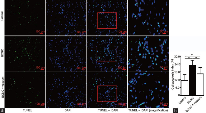
Postprostatectomy erectile dysfunction (pPED) remains a current problem despite improvements in surgical techniques. Vacuum therapy is clinically confirmed as a type of pPED rehabilitation. However, its underlying mechanisms are incompletely understood. Recently, autophagy and apoptosis were extensively studied in erectile dysfunction resulting from diabetes, senescence, and androgen deprivation but not in the context of pPED and vacuum therapy. Therefore, this study was designed to investigate the roles of autophagy and apoptosis in pPED and vacuum therapy. Twenty-four adult male Sprague-Dawley rats were randomly divided into three groups: the control group, bilateral cavernous nerve crush (BCNC) group, and BCNC + vacuum group. After 4 weeks of treatment, intracavernosal pressure was used to evaluate erectile function. Real-time quantitative polymerase chain reaction, western blot, and immunohistochemistry were used to measure the molecular expression. TdT-mediated dUTP nick-end labeling staining was used to assess apoptosis. Transmission electron microscopy was used to observe autophagosomes. After treatment, compared with those of the BCNC group, erectile function and cavernosal hypoxia had statistically significantly improved (P < 0.05). Apoptosis and the relative protein expression of B-cell lymphoma-2-associated X and cleaved Caspase3 were decreased (P < 0.05). Autophagy-related molecules such as phosphorylated unc-51-like autophagy-activating kinase 1 (Ser757) and p62 were decreased. Beclin1, microtubule-associated protein 1 light chain 3 A/B, and autophagosomes were increased (P < 0.05). Besides, the phosphatidylinositol 3-kinase/AKT/mammalian target of rapamycin signaling pathway, as a negative regulator of autophagy to some degree, was inhibited. This study revealed that vacuum therapy ameliorated pPED in BCNC rats by inhibiting apoptosis and activating autophagy.
前列腺切除术后勃起功能障碍(pPED)尽管手术技术有所提高,但仍是一个当前的问题。真空疗法在临床上被确认为 pPED 康复的一种类型。然而,其潜在机制尚不完全清楚。最近,自噬和细胞凋亡在糖尿病、衰老和雄激素剥夺引起的勃起功能障碍中得到了广泛研究,但在 pPED 和真空治疗的背景下尚未得到研究。因此,本研究旨在探讨自噬和细胞凋亡在 pPED 和真空治疗中的作用。24 只成年雄性 Sprague-Dawley 大鼠随机分为三组:对照组、双侧海绵体神经挤压(BCNC)组和 BCNC+真空组。治疗 4 周后,采用海绵体内压评估勃起功能。实时定量聚合酶链反应、Western blot 和免疫组织化学法用于测量分子表达。TdT 介导的 dUTP 缺口末端标记染色用于评估细胞凋亡。透射电子显微镜用于观察自噬体。治疗后,与 BCNC 组相比,勃起功能和海绵体缺氧均有统计学显著改善(P<0.05)。细胞凋亡和 B 细胞淋巴瘤-2 相关 X 蛋白和裂解 Caspase3 的相对蛋白表达减少(P<0.05)。自噬相关分子如磷酸化的非典型蛋白激酶 1(Ser757)和 p62 减少。Beclin1、微管相关蛋白 1 轻链 3A/B 和自噬体增加(P<0.05)。此外,磷酸肌醇 3-激酶/AKT/雷帕霉素靶蛋白信号通路作为自噬的负调节因子,在一定程度上被抑制。本研究表明,真空疗法通过抑制细胞凋亡和激活自噬来改善 BCNC 大鼠的 pPED。