Zhao Changbo, Chen Weibo, Gong Fangxin, Wang Wenzhen, Fan Cuiqin, Li Ye, Lan Tian, Wang Wenjing, Yuan Mingzhen
Department of Urology, Shandong Provincial Hospital, Shandong University, Jinan, China.
Department of Andrology, Liaocheng People's Hospital, Shandong University, Liaocheng, China.
Transl Androl Urol. 2024 Apr 30;13(4):537-547. doi: 10.21037/tau-23-547. Epub 2024 Apr 9.
Inflammation, fibrosis and autophagy represent closely related factors associated with the pathogenesis of diabetes mellitus erectile dysfunction (DMED). In this study, the therapeutic effect of nitro-oleic acid (NO-OA) in a streptozotocin-induced rat model of DMED was evaluated.
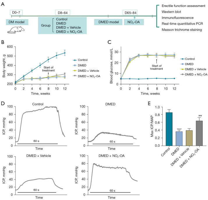
Sixty rats were randomly divided into four groups: control, DMED, DMED + Vehicle and DMED + NO-OA. DMED was induced by intraperitoneal injection of streptozotocin in male rats. Blood glucose and body weight were measured every 2 weeks. After 4 weeks of NO-OA treatment, erectile function was measured by electrical stimulation of cavernous nerve (CN). Western blotting, quantitative real-time polymerase chain reaction (qRT-PCR), enzyme-linked immunosorbent assay (ELISA), immunofluorescence and Masson's trichrome staining were used to verify the related factors and protein expression levels.
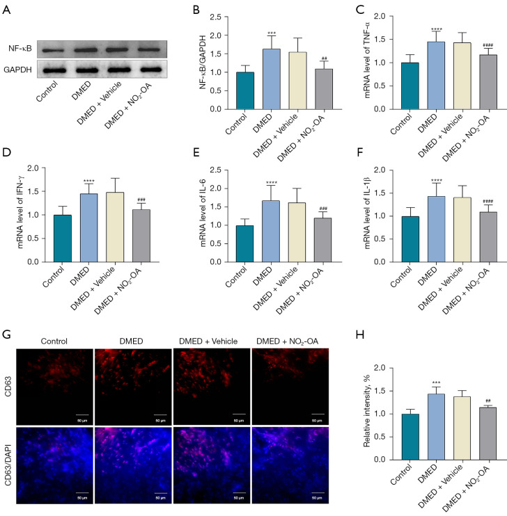
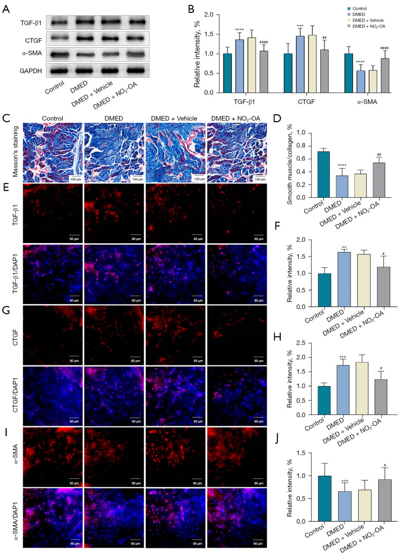
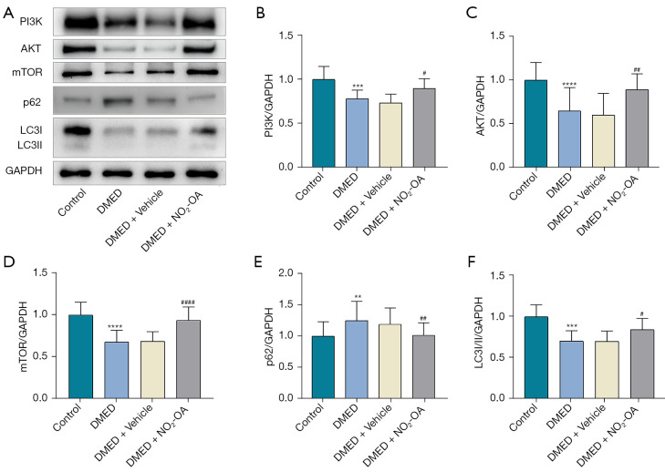
We found that NO-OA could significantly increase erectile pressure in the corpus cavernosum of DMED rats. Results of western blot, confocal immunofluorescence and qRT-PCR assays revealed that NO-OA significantly reduced inflammatory factors and the expression of nuclear factor kappa B (NF-κB). In addition, Masson staining results indicated that NO-OA significantly reduced the display of fibrotic tissue in the corpus cavernosum. These beneficial effects may be related to reductions in the expression of transforming growth factor-β1 (TGF-β1) and connective tissue growth factor (CTGF) and the increase in the expression of α-smooth muscle actin (α-SMA). Finally, NO-OA treatment increased the expression of the autophagy marker, LC3, while P62 was decreased, effects suggesting that one of the underlying mechanisms of NO-OA may involve an activation of the PI3K/AKT/mTOR pathway to enhance the capacity for autophagy within this tissue.
NO-OA enhances erectile function within a rat model of DMED by inhibiting inflammation and fibrosis along with activating autophagy.
炎症、纤维化和自噬是与糖尿病性勃起功能障碍(DMED)发病机制密切相关的因素。在本研究中,评估了硝基油酸(NO-OA)对链脲佐菌素诱导的DMED大鼠模型的治疗效果。
将60只大鼠随机分为四组:对照组、DMED组、DMED + 赋形剂组和DMED + NO-OA组。通过腹腔注射链脲佐菌素诱导雄性大鼠发生DMED。每2周测量血糖和体重。NO-OA治疗4周后,通过海绵体神经(CN)电刺激测量勃起功能。采用蛋白质免疫印迹法、定量实时聚合酶链反应(qRT-PCR)、酶联免疫吸附测定(ELISA)、免疫荧光和Masson三色染色法来验证相关因子和蛋白表达水平。
我们发现NO-OA可显著增加DMED大鼠海绵体的勃起压力。蛋白质免疫印迹、共聚焦免疫荧光和qRT-PCR检测结果显示,NO-OA可显著降低炎症因子及核因子κB(NF-κB)的表达。此外,Masson染色结果表明,NO-OA可显著减少海绵体中纤维化组织的表现。这些有益作用可能与转化生长因子-β1(TGF-β1)和结缔组织生长因子(CTGF)表达的降低以及α-平滑肌肌动蛋白(α-SMA)表达的增加有关。最后,NO-OA治疗增加了自噬标志物LC3的表达,而P62减少,这些结果表明NO-OA的潜在机制之一可能涉及激活PI3K/AKT/mTOR途径以增强该组织内的自噬能力。
NO-OA通过抑制炎症和纤维化以及激活自噬来增强DMED大鼠模型的勃起功能。