• 文献检索
  • 文档翻译
  • 深度研究
  • 学术资讯
  • Suppr Zotero 插件Zotero 插件
  • 邀请有礼
  • 套餐&价格
  • 历史记录
应用&插件
Suppr Zotero 插件Zotero 插件浏览器插件Mac 客户端Windows 客户端微信小程序
定价
高级版会员购买积分包购买API积分包
服务
文献检索文档翻译深度研究API 文档MCP 服务
关于我们
关于 Suppr公司介绍联系我们用户协议隐私条款
关注我们

Suppr 超能文献

核心技术专利:CN118964589B侵权必究
粤ICP备2023148730 号-1Suppr @ 2026

文献检索

告别复杂PubMed语法,用中文像聊天一样搜索,搜遍4000万医学文献。AI智能推荐,让科研检索更轻松。

立即免费搜索

文件翻译

保留排版,准确专业,支持PDF/Word/PPT等文件格式,支持 12+语言互译。

免费翻译文档

深度研究

AI帮你快速写综述,25分钟生成高质量综述,智能提取关键信息,辅助科研写作。

立即免费体验

弥散加权 MRI 可改善 NSCLC 患者根治性放疗后的反应评估。

Diffusion-weighted MRI improves response assessment after definitive radiotherapy in patients with NSCLC.

机构信息

Clinic for Diagnostic and Interventional Radiology, Saarland University Medical Center, Kirrberger Str. 1, 66421, Homburg, Saar, Germany.

Department of Radiotherapy and Radiation Oncology, Saarland University Medical Center, Kirrberger Str. Geb. 6.5, 66421, Homburg, Saar, Germany.

出版信息

Cancer Imaging. 2021 Jan 21;21(1):15. doi: 10.1186/s40644-021-00384-9.

DOI:10.1186/s40644-021-00384-9
PMID:33478592
原文链接:https://pmc.ncbi.nlm.nih.gov/articles/PMC7818746/
Abstract

BACKGROUND

Computed tomography (CT) is the standard procedure for follow-up of non-small-cell lung cancer (NSCLC) after radiochemotherapy. CT has difficulties differentiating between tumor, atelectasis and radiation induced lung toxicity (RILT). Diffusion-weighted imaging (DWI) may enable a more accurate detection of vital tumor tissue. The aim of this study was to determine the diagnostic value of MRI versus CT in the follow-up of NSCLC.

METHODS

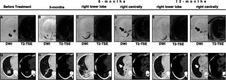
Twelve patients with NSCLC stages I-III scheduled for radiochemotherapy were enrolled in this prospective study. CT with i.v. contrast agent and non enhanced MRI were performed before and 3, 6 and 12 months after treatment. Standardized ROIs were used to determine the apparent diffusion weighted coefficient (ADC) within the tumor. Tumor size was assessed by the longest longitudinal diameter (LD) and tumor volume on DWI and CT. RILT was assessed on a 4-point-score in breath-triggered T2-TSE and CT.

RESULTS

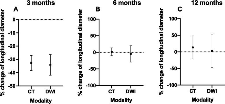
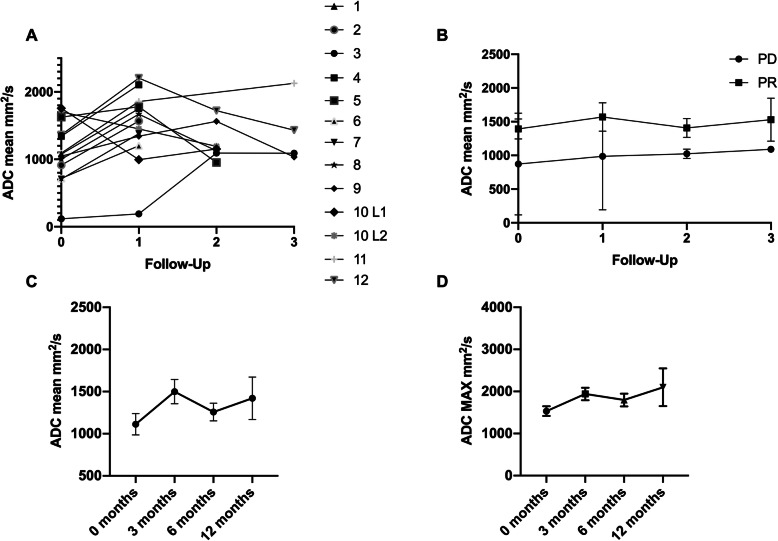
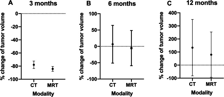
There was no significant difference regarding LD and tumor volume between MRI and CT (p ≥ 0.6221, respectively p ≥ 0.25). Evaluation of RILT showed a very high correlation between MRI and CT at 3 (r = 0.8750) and 12 months (r = 0.903). Assessment of the ADC values suggested that patients with a good tumor response have higher ADC values than non-responders.

CONCLUSIONS

DWI is equivalent to CT for tumor volume determination in patients with NSCLC during follow up. The extent of RILT can be reliably determined by MRI. DWI could become a beneficial method to assess tumor response more accurately. ADC values may be useful as a prognostic marker.

摘要

背景

计算机断层扫描(CT)是放射化学治疗后非小细胞肺癌(NSCLC)随访的标准程序。CT 难以区分肿瘤、肺不张和放射性肺毒性(RILT)。扩散加权成像(DWI)可能使肿瘤组织的检测更加准确。本研究旨在确定 MRI 与 CT 在 NSCLC 随访中的诊断价值。

方法

本前瞻性研究纳入了 12 例计划接受放射化学治疗的 NSCLC Ⅰ-Ⅲ期患者。治疗前、治疗后 3、6 和 12 个月进行 CT 增强扫描和 MRI 平扫。在肿瘤内使用标准化 ROI 确定表观扩散系数(ADC)。通过最长纵向直径(LD)和 DWI 和 CT 上的肿瘤体积评估肿瘤大小。在呼吸触发 T2-TSE 和 CT 上进行 4 分制评分评估 RILT。

结果

MRI 和 CT 之间的 LD 和肿瘤体积无显著差异(分别为 p≥0.6221 和 p≥0.25)。RILT 的评估显示 MRI 和 CT 在 3 个月(r=0.8750)和 12 个月(r=0.903)时具有非常高的相关性。ADC 值评估表明,肿瘤反应良好的患者 ADC 值高于无反应者。

结论

在 NSCLC 患者随访期间,DWI 与 CT 相比,在肿瘤体积确定方面具有等效性。RILT 的程度可以通过 MRI 可靠地确定。DWI 可能成为一种评估肿瘤反应更准确的有益方法。ADC 值可能作为一种预后标志物。

https://cdn.ncbi.nlm.nih.gov/pmc/blobs/3a12/7818746/2acf1181caaa/40644_2021_384_Fig4_HTML.jpg
https://cdn.ncbi.nlm.nih.gov/pmc/blobs/3a12/7818746/c6c24fd542ba/40644_2021_384_Fig1_HTML.jpg
https://cdn.ncbi.nlm.nih.gov/pmc/blobs/3a12/7818746/fcfeb65938a8/40644_2021_384_Fig2_HTML.jpg
https://cdn.ncbi.nlm.nih.gov/pmc/blobs/3a12/7818746/fc796a285986/40644_2021_384_Fig3_HTML.jpg
https://cdn.ncbi.nlm.nih.gov/pmc/blobs/3a12/7818746/2acf1181caaa/40644_2021_384_Fig4_HTML.jpg

相似文献

1
Diffusion-weighted MRI improves response assessment after definitive radiotherapy in patients with NSCLC.弥散加权 MRI 可改善 NSCLC 患者根治性放疗后的反应评估。
Cancer Imaging. 2021 Jan 21;21(1):15. doi: 10.1186/s40644-021-00384-9.
2
Diffusion-weighted MRI of the lung at 3T evaluated using echo-planar-based and single-shot turbo spin-echo-based acquisition techniques for radiotherapy applications.使用基于回波平面和基于单次激发快速自旋回波的采集技术,对3T磁共振成像下的肺部进行扩散加权成像,以用于放射治疗应用。
J Appl Clin Med Phys. 2019 Jan;20(1):284-292. doi: 10.1002/acm2.12493. Epub 2018 Nov 12.
3
The Impact of Diffusion-Weighted MRI on the Definition of Gross Tumor Volume in Radiotherapy of Non-Small-Cell Lung Cancer.扩散加权磁共振成像对非小细胞肺癌放疗中大体肿瘤体积定义的影响
PLoS One. 2016 Sep 9;11(9):e0162816. doi: 10.1371/journal.pone.0162816. eCollection 2016.
4
Change in Apparent Diffusion Coefficient Is Associated With Local Failure After Stereotactic Body Radiation Therapy for Non-Small Cell Lung Cancer: A Prospective Clinical Trial.在立体定向体部放射治疗非小细胞肺癌后,表观扩散系数的变化与局部失败相关:一项前瞻性临床试验。
Int J Radiat Oncol Biol Phys. 2019 Nov 1;105(3):659-663. doi: 10.1016/j.ijrobp.2019.06.2536. Epub 2019 Jul 2.
5
Apparent diffusion coefficient (ADC) change on repeated diffusion-weighted magnetic resonance imaging during radiochemotherapy for non-small cell lung cancer: A pilot study.非小细胞肺癌放化疗期间重复扩散加权磁共振成像的表观扩散系数(ADC)变化:一项初步研究。
Lung Cancer. 2016 Jun;96:113-9. doi: 10.1016/j.lungcan.2016.04.001. Epub 2016 Apr 4.
6
Diffusion-weighted MRI versus 18F-FDG PET/CT: performance as predictors of tumor treatment response and patient survival in patients with non-small cell lung cancer receiving chemoradiotherapy.弥散加权 MRI 与 18F-FDG PET/CT 对比:在接受放化疗的非小细胞肺癌患者中,预测肿瘤治疗反应和患者生存的性能。
AJR Am J Roentgenol. 2012 Jan;198(1):75-82. doi: 10.2214/AJR.11.6525.
7
Diffusion weighted MRI and 18F-FDG PET/CT in non-small cell lung cancer (NSCLC): does the apparent diffusion coefficient (ADC) correlate with tracer uptake (SUV)?弥散加权 MRI 与 18F-FDG PET/CT 在非小细胞肺癌(NSCLC)中的应用:表观扩散系数(ADC)与示踪剂摄取(SUV)是否相关?
Eur J Radiol. 2012 Oct;81(10):2913-8. doi: 10.1016/j.ejrad.2011.11.050. Epub 2011 Dec 23.
8
Hybrid [¹⁸F]-FDG PET/MRI including non-Gaussian diffusion-weighted imaging (DWI): preliminary results in non-small cell lung cancer (NSCLC).¹⁸F-氟代脱氧葡萄糖正电子发射断层扫描/磁共振成像(¹⁸F-FDG PET/MRI)联合非高斯扩散加权成像(DWI):非小细胞肺癌(NSCLC)的初步结果。
Eur J Radiol. 2013 Nov;82(11):2055-60. doi: 10.1016/j.ejrad.2013.05.027. Epub 2013 Jul 5.
9
A Prospective Study of Magnetic Resonance Imaging Assessment of Post-radiation Changes Following Stereotactic Body Radiation Therapy for Non-small Cell Lung Cancer.一项立体定向体部放射治疗非小细胞肺癌后放射性改变的磁共振成像前瞻性研究。
Clin Oncol (R Coll Radiol). 2019 Oct;31(10):720-727. doi: 10.1016/j.clon.2019.05.014. Epub 2019 Jun 5.
10
18F-FDG PET/CT and whole-body MRI diagnostic performance in M staging for non-small cell lung cancer: a systematic review and meta-analysis.18F-FDG PET/CT 和全身 MRI 在非小细胞肺癌 M 分期中的诊断性能:系统评价和荟萃分析。
Eur Radiol. 2020 Jul;30(7):3641-3649. doi: 10.1007/s00330-020-06703-1. Epub 2020 Mar 3.

引用本文的文献

1
Development and validation of multi-sequence magnetic resonance imaging radiomics models for predicting tumor response to radiotherapy in locally advanced non-small cell lung cancer.用于预测局部晚期非小细胞肺癌放疗肿瘤反应的多序列磁共振成像放射组学模型的开发与验证
J Thorac Dis. 2025 Mar 31;17(3):1684-1697. doi: 10.21037/jtd-2025-142. Epub 2025 Mar 14.
2
Radiogenomics models for predicting prognosis in locally advanced non-small cell lung cancer patients undergoing definitive chemoradiotherapy.用于预测接受根治性放化疗的局部晚期非小细胞肺癌患者预后的放射基因组学模型。
Transl Lung Cancer Res. 2024 Aug 31;13(8):1828-1840. doi: 10.21037/tlcr-24-145. Epub 2024 Aug 28.
3
Effectiveness of Apparent Diffusion Coefficient Values in Predicting Pathologic Subtypes and Grade in Non-Small-Cell Lung Cancer.
表观扩散系数值在预测非小细胞肺癌病理亚型和分级中的有效性
Diagnostics (Basel). 2024 Aug 16;14(16):1795. doi: 10.3390/diagnostics14161795.
4
Delta-radiomics features of ADC maps as early predictors of treatment response in lung cancer.作为肺癌治疗反应早期预测指标的ADC图的Delta放射组学特征
Insights Imaging. 2024 Aug 26;15(1):218. doi: 10.1186/s13244-024-01787-5.
5
Histogram analysis of multiple diffusion models for predicting advanced non-small cell lung cancer response to chemoimmunotherapy.多扩散模型直方图分析预测晚期非小细胞肺癌对化疗免疫治疗的反应。
Cancer Imaging. 2024 Jun 11;24(1):71. doi: 10.1186/s40644-024-00713-8.
6
New developments in the imaging of lung cancer.肺癌成像的新进展。
Breathe (Sheff). 2024 Mar;20(1):230176. doi: 10.1183/20734735.0176-2023. Epub 2024 Apr 9.
7
The lung cancers: staging and response, CT, F-FDG PET/CT, MRI, DWI: review and new perspectives.肺癌的分期和疗效评价:CT、F-FDG PET/CT、MRI、DWI——综述及新视角
Br J Radiol. 2023 Aug;96(1148):20220339. doi: 10.1259/bjr.20220339. Epub 2023 May 17.
8
Repeatability and test-retest reproducibility of mean apparent diffusion coefficient measurements of focal and diffuse disease in relapsed multiple myeloma at 3T whole body diffusion-weighted MRI (WB-DW-MRI).在 3T 全身扩散加权 MRI(WB-DW-MRI)中,复发性多发性骨髓瘤的局灶性和弥漫性病变的平均表观扩散系数测量的重复性和测试-再测试可重复性。
Br J Radiol. 2022 Sep 1;95(1138):20220418. doi: 10.1259/bjr.20220418. Epub 2022 Jul 28.
9
CT Combined with Multiparameter MRI in Differentiating Pathological Subtypes of Non-Small-Cell Lung Cancer before Surgery.CT 联合多参数 MRI 术前鉴别非小细胞肺癌病理亚型。
Contrast Media Mol Imaging. 2022 May 17;2022:8207301. doi: 10.1155/2022/8207301. eCollection 2022.
10
MR-guided radiotherapy in node-positive non-small cell lung cancer and severely limited pulmonary reserve: a report proposing a new clinical pathway for the management of high-risk patients.MR 引导放疗在淋巴结阳性非小细胞肺癌和严重肺储备功能受限患者中的应用:报告提出一种管理高危患者的新临床路径。
Radiat Oncol. 2022 Feb 24;17(1):43. doi: 10.1186/s13014-022-02011-8.