Pulmonary Biology, The Perinatal Institute and Section of Neonatology, Cincinnati Children's Hospital Medical Center, Cincinnati, Ohio, USA.
Division of Pulmonary and Critical Care Medicine, Harvard Medical School, Boston, Massachusetts, USA.
Thorax. 2021 May;76(5):456-467. doi: 10.1136/thoraxjnl-2020-214986. Epub 2021 Jan 21.
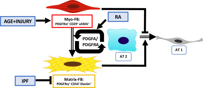
Idiopathic pulmonary fibrosis (IPF) primarily affects the aged population and is characterised by failure of alveolar regeneration, leading to loss of alveolar type 1 (AT1) cells. Aged mouse models of lung repair have demonstrated that regeneration fails with increased age. Mouse and rat lung repair models have shown retinoic acid (RA) treatment can restore alveolar regeneration. Herein, we seek to determine the signalling mechanisms that become activated on RA treatment prior to injury, which support alveolar differentiation.
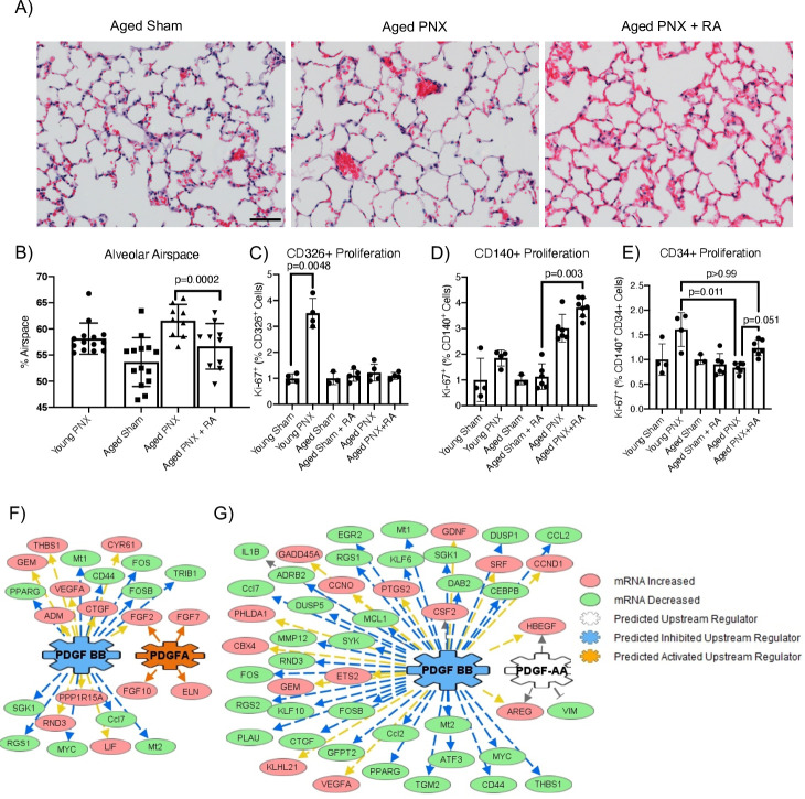
Partial pneumonectomy lung injury model and next-generation sequencing of sorted cell populations were used to uncover molecular targets regulating alveolar repair. In vitro organoids generated from epithelial cells of mouse or patient with IPF co-cultured with young, aged or RA-pretreated murine fibroblasts were used to test potential targets.
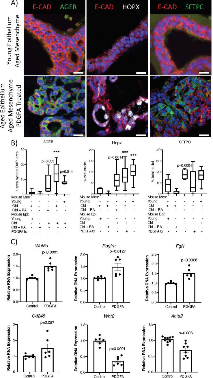
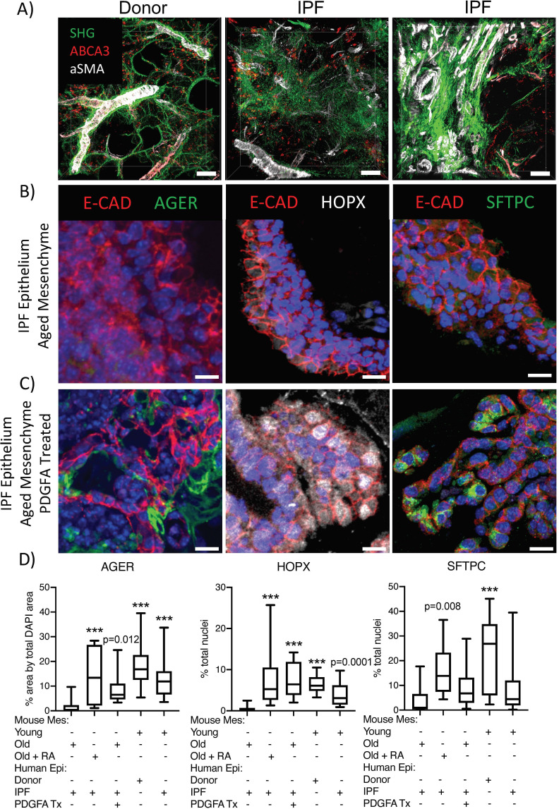
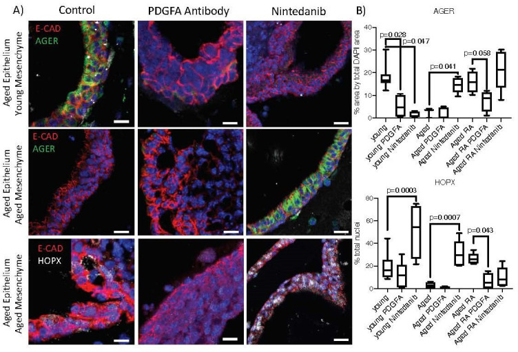
Known alveolar epithelial cell differentiation markers, including HOPX and AGER for AT1 cells, were used to assess outcome of treatments.
Gene expression analysis of sorted fibroblasts and epithelial cells isolated from lungs of young, aged and RA-pretreated aged mice predicted increased platelet-derived growth factor subunit A (PDGFA) signalling that coincided with regeneration and alveolar epithelial differentiation. Addition of PDGFA induced AT1 and AT2 differentiation in both mouse and human IPF lung organoids generated with aged fibroblasts, and PDGFA monoclonal antibody blocked AT1 cell differentiation in organoids generated with young murine fibroblasts.
Our data support the concept that RA indirectly induces reciprocal PDGFA signalling, which activates regenerative fibroblasts that support alveolar epithelial cell differentiation and repair, providing a potential therapeutic strategy to influence the pathogenesis of IPF.
特发性肺纤维化(IPF)主要影响老年人群,其特征是肺泡再生失败,导致肺泡 1 型(AT1)细胞丧失。修复肺的老年小鼠模型表明,随着年龄的增长,再生会失败。鼠和大鼠肺修复模型表明,视黄酸(RA)治疗可以恢复肺泡再生。在此,我们试图确定在损伤前 RA 治疗激活的信号转导机制,这些机制支持肺泡分化。
采用部分肺切除术肺损伤模型和分选细胞群体的下一代测序技术,揭示调节肺泡修复的分子靶标。从 IPF 患者的上皮细胞和小鼠中生成的类器官与年轻、老年或 RA 预处理的鼠成纤维细胞共培养,用于测试潜在的靶标。
已知的肺泡上皮细胞分化标志物,包括 AT1 细胞的 HOPX 和AGER,用于评估治疗结果。
从小鼠肺中分离的分选成纤维细胞和上皮细胞的基因表达分析预测,血小板衍生生长因子亚单位 A(PDGFA)信号转导增加,这与再生和肺泡上皮细胞分化相一致。在使用老年成纤维细胞生成的鼠和人 IPF 肺类器官中,添加 PDGFA 可诱导 AT1 和 AT2 分化,而 PDGFA 单克隆抗体可阻止使用年轻鼠成纤维细胞生成的类器官中 AT1 细胞分化。
我们的数据支持这样的概念,即 RA 间接诱导 PDGFA 信号的相互作用,这种相互作用激活了支持肺泡上皮细胞分化和修复的再生成纤维细胞,为影响 IPF 的发病机制提供了一种潜在的治疗策略。