• 文献检索
  • 文档翻译
  • 深度研究
  • 学术资讯
  • Suppr Zotero 插件Zotero 插件
  • 邀请有礼
  • 套餐&价格
  • 历史记录
应用&插件
Suppr Zotero 插件Zotero 插件浏览器插件Mac 客户端Windows 客户端微信小程序
定价
高级版会员购买积分包购买API积分包
服务
文献检索文档翻译深度研究API 文档MCP 服务
关于我们
关于 Suppr公司介绍联系我们用户协议隐私条款
关注我们

Suppr 超能文献

核心技术专利:CN118964589B侵权必究
粤ICP备2023148730 号-1Suppr @ 2026

文献检索

告别复杂PubMed语法,用中文像聊天一样搜索,搜遍4000万医学文献。AI智能推荐,让科研检索更轻松。

立即免费搜索

文件翻译

保留排版,准确专业,支持PDF/Word/PPT等文件格式,支持 12+语言互译。

免费翻译文档

深度研究

AI帮你快速写综述,25分钟生成高质量综述,智能提取关键信息,辅助科研写作。

立即免费体验

左心房结扎驱动的异常血流动力学对胚胎心脏发育的影响:左心发育不全综合征产前进展的线索。

Effect of left atrial ligation-driven altered inflow hemodynamics on embryonic heart development: clues for prenatal progression of hypoplastic left heart syndrome.

机构信息

Biomedical Research Center, Qatar University, Doha, Qatar.

Department of Mechanical Engineering, TOBB University of Economics and Technology, Ankara, Turkey.

出版信息

Biomech Model Mechanobiol. 2021 Apr;20(2):733-750. doi: 10.1007/s10237-020-01413-5. Epub 2021 Jan 22.

DOI:10.1007/s10237-020-01413-5
PMID:33481120
原文链接:https://pmc.ncbi.nlm.nih.gov/articles/PMC7979615/
Abstract

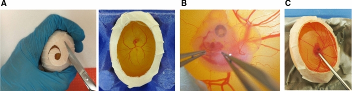
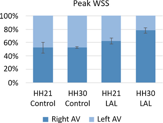
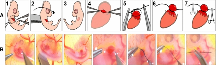
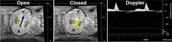
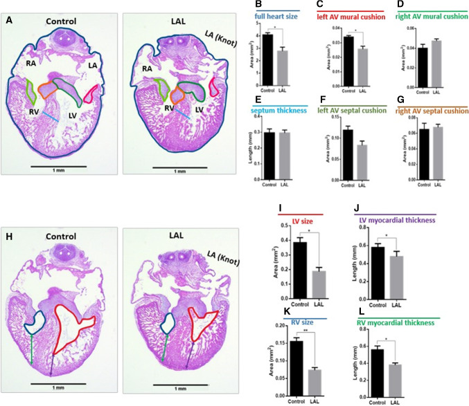
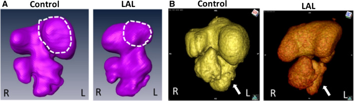
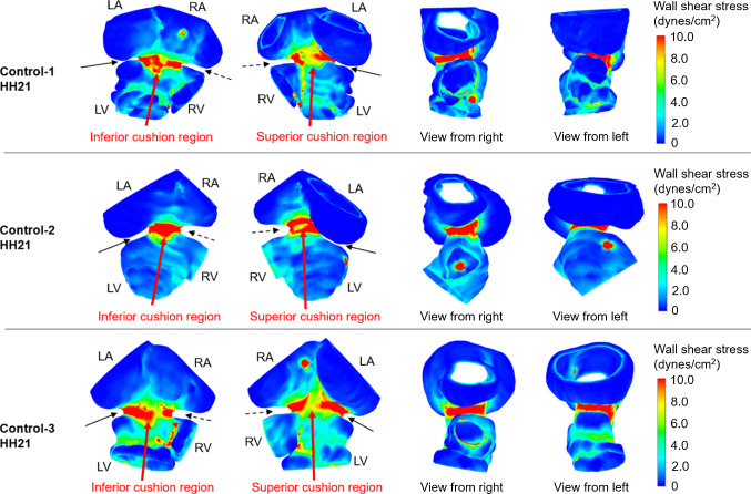
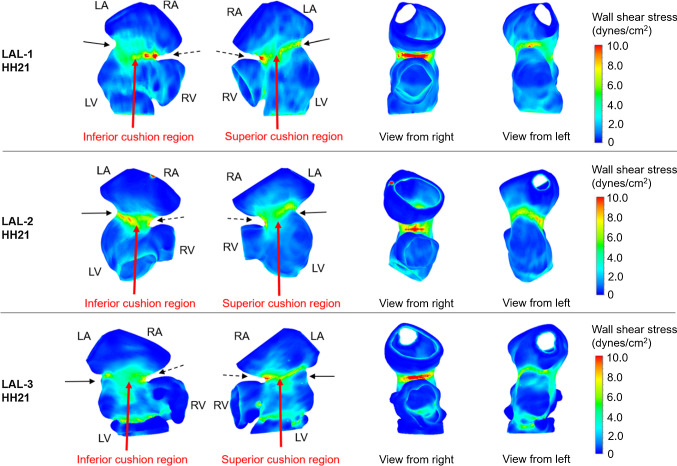
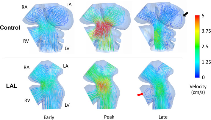
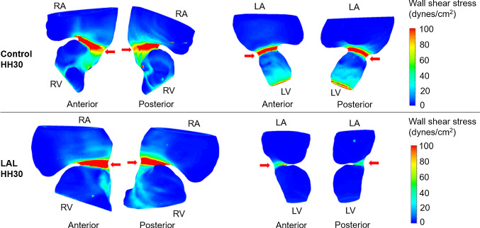
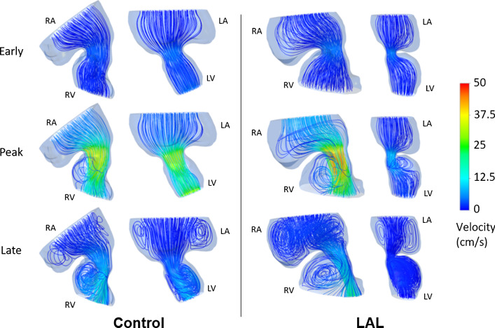
Congenital heart defects (CHDs) are abnormalities in the heart structure present at birth. One important condition is hypoplastic left heart syndrome (HLHS) where severely underdeveloped left ventricle (LV) cannot support systemic circulation. HLHS usually initiates as localized tissue malformations with no underlying genetic cause, suggesting that disturbed hemodynamics contribute to the embryonic development of these defects. Left atrial ligation (LAL) is a surgical procedure on embryonic chick resulting in a phenotype resembling clinical HLHS. In this study, we investigated disturbed hemodynamics and deteriorated cardiac growth following LAL to investigate possible mechanobiological mechanisms for the embryonic development of HLHS. We integrated techniques such as echocardiography, micro-CT and computational fluid dynamics (CFD) for these analyses. Specifically, LAL procedure causes an immediate flow disturbance over atrioventricular (AV) cushions. At later stages after the heart septation, it causes hemodynamic disturbances in LV. As a consequence of the LAL procedure, the left-AV canal and LV volume decrease in size, and in the opposite way, the right-AV canal and right ventricle volume increase. According to our CFD analysis, LAL results in an immediate decrease in the left AV canal WSS levels for 3.5-day (HH21) pre-septated hearts. For 7-day post-septated hearts (HH30), LAL leads to further reduction in WSS levels in the left AV canal, and relatively increased WSS levels in the right AV canal. This study demonstrates the critical importance of the disturbed hemodynamics during the heart valve and ventricle development.

摘要

先天性心脏缺陷(CHD)是出生时存在的心脏结构异常。一种重要的情况是左心发育不全综合征(HLHS),其中严重发育不良的左心室(LV)无法支持全身循环。HLHS 通常最初表现为局部组织畸形,没有潜在的遗传原因,这表明血流动力学紊乱有助于这些缺陷的胚胎发育。左心房结扎(LAL)是对胚胎鸡进行的一种手术,导致类似于临床 HLHS 的表型。在这项研究中,我们研究了 LAL 后血流动力学紊乱和心脏生长恶化的情况,以研究 HLHS 胚胎发育的可能机械生物学机制。我们整合了超声心动图、微 CT 和计算流体动力学(CFD)等技术进行这些分析。具体来说,LAL 手术会立即引起房室(AV)瓣垫的血流紊乱。在心脏分隔后期,它会导致 LV 的血流动力学紊乱。由于 LAL 手术,左 AV 瓣口和 LV 容积减小,而右 AV 瓣口和右心室容积增大。根据我们的 CFD 分析,LAL 会导致未分隔心脏(HH21)的左 AV 瓣口 WSS 水平立即下降。对于分隔后 7 天(HH30)的心脏,LAL 导致左 AV 瓣口的 WSS 水平进一步降低,而右 AV 瓣口的 WSS 水平相对增加。这项研究表明,在心脏瓣膜和心室发育过程中,血流动力学紊乱至关重要。

https://cdn.ncbi.nlm.nih.gov/pmc/blobs/e908/7979615/06e800262679/10237_2020_1413_Fig16_HTML.jpg
https://cdn.ncbi.nlm.nih.gov/pmc/blobs/e908/7979615/0bb11cab4342/10237_2020_1413_Fig1_HTML.jpg
https://cdn.ncbi.nlm.nih.gov/pmc/blobs/e908/7979615/1b6d523ca8fa/10237_2020_1413_Fig2_HTML.jpg
https://cdn.ncbi.nlm.nih.gov/pmc/blobs/e908/7979615/3d537cc8366a/10237_2020_1413_Fig3_HTML.jpg
https://cdn.ncbi.nlm.nih.gov/pmc/blobs/e908/7979615/e2232608d290/10237_2020_1413_Fig4_HTML.jpg
https://cdn.ncbi.nlm.nih.gov/pmc/blobs/e908/7979615/c72922f12621/10237_2020_1413_Fig5_HTML.jpg
https://cdn.ncbi.nlm.nih.gov/pmc/blobs/e908/7979615/7608f20e71ee/10237_2020_1413_Fig6_HTML.jpg
https://cdn.ncbi.nlm.nih.gov/pmc/blobs/e908/7979615/1eb52fbad70a/10237_2020_1413_Fig7_HTML.jpg
https://cdn.ncbi.nlm.nih.gov/pmc/blobs/e908/7979615/8c35bcb35e94/10237_2020_1413_Fig8_HTML.jpg
https://cdn.ncbi.nlm.nih.gov/pmc/blobs/e908/7979615/7592ce9acda8/10237_2020_1413_Fig9_HTML.jpg
https://cdn.ncbi.nlm.nih.gov/pmc/blobs/e908/7979615/ac5e6b5114b0/10237_2020_1413_Fig10_HTML.jpg
https://cdn.ncbi.nlm.nih.gov/pmc/blobs/e908/7979615/9581b68bd10f/10237_2020_1413_Fig11_HTML.jpg
https://cdn.ncbi.nlm.nih.gov/pmc/blobs/e908/7979615/0855aeb8352c/10237_2020_1413_Fig12_HTML.jpg
https://cdn.ncbi.nlm.nih.gov/pmc/blobs/e908/7979615/341b404bc672/10237_2020_1413_Fig13_HTML.jpg
https://cdn.ncbi.nlm.nih.gov/pmc/blobs/e908/7979615/460630611161/10237_2020_1413_Fig14_HTML.jpg
https://cdn.ncbi.nlm.nih.gov/pmc/blobs/e908/7979615/1946b4905965/10237_2020_1413_Fig15_HTML.jpg
https://cdn.ncbi.nlm.nih.gov/pmc/blobs/e908/7979615/06e800262679/10237_2020_1413_Fig16_HTML.jpg

相似文献

1
Effect of left atrial ligation-driven altered inflow hemodynamics on embryonic heart development: clues for prenatal progression of hypoplastic left heart syndrome.左心房结扎驱动的异常血流动力学对胚胎心脏发育的影响:左心发育不全综合征产前进展的线索。
Biomech Model Mechanobiol. 2021 Apr;20(2):733-750. doi: 10.1007/s10237-020-01413-5. Epub 2021 Jan 22.
2
Fluid mechanics of the left atrial ligation chick embryonic model of hypoplastic left heart syndrome.左心房结扎雏鸡左心发育不全综合征模型的流体力学。
Biomech Model Mechanobiol. 2021 Aug;20(4):1337-1351. doi: 10.1007/s10237-021-01447-3. Epub 2021 Mar 28.
3
Left atrial ligation alters intracardiac flow patterns and the biomechanical landscape in the chick embryo.左心房结扎改变了鸡胚的心内血流模式和生物力学环境。
Dev Dyn. 2014 May;243(5):652-62. doi: 10.1002/dvdy.24107.
4
Myocardial Biomechanics and the Consequent Differentially Expressed Genes of the Left Atrial Ligation Chick Embryonic Model of Hypoplastic Left Heart Syndrome.左心房结扎 Chick 胚胎模型的左心发育不全综合征心肌生物力学与差异表达基因。
Ann Biomed Eng. 2023 May;51(5):1063-1078. doi: 10.1007/s10439-023-03187-0. Epub 2023 Apr 9.
5
Blood Flow Disturbance and Morphological Alterations Following the Right Atrial Ligation in the Chick Embryo.鸡胚右心房结扎后的血流紊乱与形态学改变
Front Physiol. 2022 Apr 14;13:849603. doi: 10.3389/fphys.2022.849603. eCollection 2022.
6
Right and left ventricular wall deformation patterns in normal and left heart hypoplasia chick embryos.正常和左心发育不全鸡胚的左右心室壁变形模式
Am J Physiol Heart Circ Physiol. 2000 Sep;279(3):H959-69. doi: 10.1152/ajpheart.2000.279.3.H959.
7
Right ventricle in hypoplastic left heart syndrome exhibits altered hemodynamics in the human fetus.左心发育不良综合征患者右心室在胎儿期存在血液动力学改变。
J Biomech. 2020 Nov 9;112:110035. doi: 10.1016/j.jbiomech.2020.110035. Epub 2020 Sep 6.
8
Left Atrial Ligation in the Avian Embryo as a Model for Altered Hemodynamic Loading During Early Vascular Development.左心房结扎术在禽类胚胎中作为一种改变早期血管发育期间血流动力学负荷的模型。
J Vis Exp. 2023 Jun 16(196). doi: 10.3791/65330.
9
Hemodynamic and Structural Comparison of Human Fetal Heart Development Between Normally Growing and Hypoplastic Left Heart Syndrome-Diagnosed Hearts.正常生长胎儿心脏与诊断为左心发育不全综合征胎儿心脏的血流动力学及结构比较
Front Physiol. 2022 Mar 23;13:856879. doi: 10.3389/fphys.2022.856879. eCollection 2022.
10
Energy loss and coronary flow simulation following hybrid stage I palliation: a hypoplastic left heart computational fluid dynamic model.一期杂交姑息治疗后的能量损失与冠状动脉血流模拟:一种左心发育不全的计算流体动力学模型
Interact Cardiovasc Thorac Surg. 2013 Aug;17(2):308-13. doi: 10.1093/icvts/ivt193. Epub 2013 May 9.

引用本文的文献

1
An anatomically informed computational fluid dynamics modeling approach for quantifying hemodynamics in the developing heart.一种基于解剖学信息的计算流体动力学建模方法,用于量化发育中心脏的血流动力学。
PLoS One. 2025 May 19;20(5):e0322233. doi: 10.1371/journal.pone.0322233. eCollection 2025.
2
Developmental and Evolutionary Heart Adaptations Through Structure-Function Relationships.通过结构-功能关系实现的发育和进化心脏适应
J Cardiovasc Dev Dis. 2025 Feb 22;12(3):83. doi: 10.3390/jcdd12030083.
3
Spontaneously Corrected Hypoplastic Left Heart: A Case Report and Exceptional Opportunity to Discuss Etiology with Novel Therapeutic Vision.
自发性矫正左心发育不良:病例报告及具有新治疗视角探讨病因的特殊机会。
Arch Iran Med. 2024 Sep 1;27(9):527-529. doi: 10.34172/aim.31185.
4
An optimized CT-dense agent perfusion and micro-CT imaging protocol for chick embryo developmental stages.一种针对鸡胚发育阶段的优化CT造影剂灌注和微型CT成像方案。
BMC Biomed Eng. 2024 Apr 23;6(1):3. doi: 10.1186/s42490-024-00078-w.
5
Recasting Current Knowledge of Human Fetal Circulation: The Importance of Computational Models.重塑当前关于人类胎儿循环的知识:计算模型的重要性。
J Cardiovasc Dev Dis. 2023 May 30;10(6):240. doi: 10.3390/jcdd10060240.
6
Shear and hydrostatic stress regulate fetal heart valve remodeling through YAP-mediated mechanotransduction.切应力和流体静压通过 YAP 介导体细胞机械转导调控胎儿心脏瓣膜重塑。
Elife. 2023 Apr 20;12:e83209. doi: 10.7554/eLife.83209.
7
Myocardial Biomechanics and the Consequent Differentially Expressed Genes of the Left Atrial Ligation Chick Embryonic Model of Hypoplastic Left Heart Syndrome.左心房结扎 Chick 胚胎模型的左心发育不全综合征心肌生物力学与差异表达基因。
Ann Biomed Eng. 2023 May;51(5):1063-1078. doi: 10.1007/s10439-023-03187-0. Epub 2023 Apr 9.
8
Recent advances in quantifying the mechanobiology of cardiac development via computational modeling.通过计算建模量化心脏发育机械生物学的最新进展。
Curr Opin Biomed Eng. 2023 Mar;25. doi: 10.1016/j.cobme.2022.100428. Epub 2022 Nov 21.
9
Effect of Blood Flow on Cardiac Morphogenesis and Formation of Congenital Heart Defects.血流对心脏形态发生及先天性心脏缺陷形成的影响。
J Cardiovasc Dev Dis. 2022 Sep 8;9(9):303. doi: 10.3390/jcdd9090303.
10
Flow-Mediated Factors in the Pathogenesis of Hypoplastic Left Heart Syndrome.左心发育不全综合征发病机制中的血流介导因素
J Cardiovasc Dev Dis. 2022 May 12;9(5):154. doi: 10.3390/jcdd9050154.