Clinic School of Medicine and Affiliated Hospital, North China University of Science and Technology, Tangshan 063000, China.
Hebei Key Laboratory for Chronic Diseases, North China University of Science and Technology, Tangshan 063000, China.
Oxid Med Cell Longev. 2020 Dec 31;2020:9782062. doi: 10.1155/2020/9782062. eCollection 2020.
Astragaloside IV shows neuroprotective activity, but its mechanism remains unclear. To investigate whether astragaloside IV protects from endoplasmic reticulum stress (ERS), we focus on the regulation of glycogen synthase kinase-3 (GSK-3) and mitochondrial permeability transition pore (mPTP) by astragaloside IV in neuronal cell PC12.
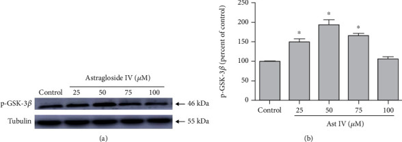
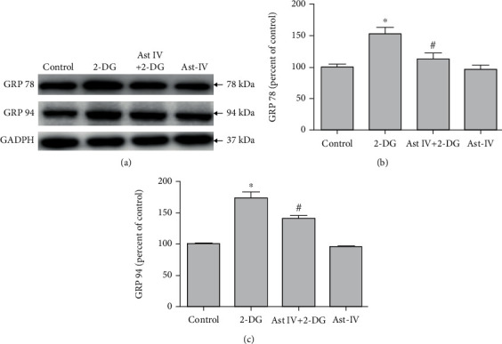
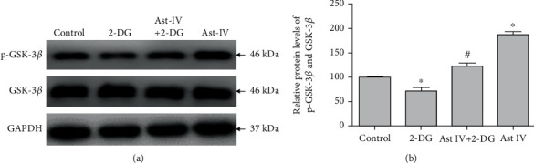
PC12 cells treated with different concentrations of ERS inductor 2-deoxyglucose (2-DG) (25-500 M) showed a significant increase of glucose-regulated protein 78 (GRP 78) and GRP 94 expressions and a decrease of tetramethylrhodamine ethyl ester (TMRE) fluorescence intensity and mitochondrial membrane potential (∆m), with the peak effect seen at 50 M, indicating that 2-DG induces ERS and the mPTP opening. Similarly, 50 M of astragaloside IV increased the GSK-3 phosphorylation at Ser9 most significantly. Next, we examined the neuroprotection of astragaloside IV by dividing the PC12 cells into control group, 2-DG treatment group, astragaloside IV plus 2-DG treatment group, and astragaloside IV only group. PC12 cells treated with 50 M 2-DG for different time courses (0-36 hr) showed a significant increase of Cleaved-Caspase-3 with the peak at 6 hr. 2-DG significantly induced cell apoptosis and increased the green fluorescence intensity of Annexin V-FITC, and these effects were reversed by astragaloside IV. Such a result indicates that astragaloside IV protected neural cell survival from ERS. 2-DG treatment significantly increased the expressions of inositol-requiring ER-to-nucleus signal kinase 1 (IRE1), phosphor-protein kinase R-like ER kinase (p-PERK), but not affect the transcription factor 6 (ATF6) expression. 2-DG treatment significantly decreased the phosphorylation of GSK-3 and significantly reduced the TMRE fluorescence intensity and ∆m, following mPTP open. Astragaloside IV significantly inhibited the above effects caused by 2-DG, except the upregulation of ATF6 protein. Taken together, astragaloside IV significantly inhibited the ERS caused by 2-DG.
Our data suggested that astragaloside IV protects PC12 cells from ERS by inactivation of GSK-3 and preventing the mPTP opening. The GRP 78, GRP 94, IRE1, and PERK signaling pathways but not ATF6 are responsible for GSK-3 inactivation and neuroprotection by astragaloside IV.
黄芪甲苷具有神经保护活性,但作用机制尚不清楚。本研究旨在探讨黄芪甲苷是否通过调节糖原合成酶激酶-3(GSK-3)和线粒体通透性转换孔(mPTP)来发挥抗内质网应激(ERS)作用,我们重点研究了黄芪甲苷对 PC12 神经元细胞的作用。
用不同浓度的 ERS 诱导剂 2-脱氧葡萄糖(2-DG)(25-500μM)处理 PC12 细胞后,葡萄糖调节蛋白 78(GRP 78)和 GRP 94 的表达明显增加,四甲基罗丹明乙酯(TMRE)荧光强度和线粒体膜电位(∆m)明显降低,在 50μM 时达到峰值,表明 2-DG 诱导 ERS 和 mPTP 开放。同样,50μM 黄芪甲苷最显著地增加了 GSK-3 丝氨酸 9 位的磷酸化。接下来,我们将 PC12 细胞分为对照组、2-DG 处理组、黄芪甲苷加 2-DG 处理组和黄芪甲苷单独处理组,通过观察不同时间点(0-36 小时)2-DG 对细胞的作用,探讨黄芪甲苷的神经保护作用。结果发现,50μM 2-DG 处理不同时间(0-36 小时)后,Cleaved-Caspase-3 明显增加,6 小时时达到峰值。2-DG 显著诱导细胞凋亡,并增加 Annexin V-FITC 的绿色荧光强度,而这些作用被黄芪甲苷逆转。这表明黄芪甲苷可以保护神经细胞免受 ERS 的影响。2-DG 处理明显增加了肌醇需求 ER 到核信号激酶 1(IRE1)和磷酸化蛋白激酶 R 样内质网激酶(p-PERK)的表达,但不影响转录因子 6(ATF6)的表达。2-DG 处理明显降低了 GSK-3 的磷酸化水平,显著降低了 TMRE 荧光强度和 ∆m,随后 mPTP 开放。黄芪甲苷显著抑制了 2-DG 引起的上述作用,除了上调 ATF6 蛋白。综上所述,黄芪甲苷通过抑制 GSK-3 的活性和防止 mPTP 的开放来抑制 2-DG 引起的 ERS。葡萄糖调节蛋白 78、94、IRE1 和 PERK 信号通路而不是 ATF6 负责黄芪甲苷抑制 GSK-3 活性和发挥神经保护作用。