Duprez Jessica, Kalbfleisch Kristen, Deshmukh Sasmit, Payne Jessie, Haer Manjit, Williams Wayne, Durowoju Ibrahim, Kirkitadze Marina
Analytical Sciences, Sanofi Pasteur Canada, 1755 Steeles Avenue West, Toronto, Ontario, Canada.
Department of Biology, York University, 4700 Keele Street, Toronto, Ontario, Canada.
Comput Struct Biotechnol J. 2020 Dec 23;19:439-447. doi: 10.1016/j.csbj.2020.12.023. eCollection 2021.
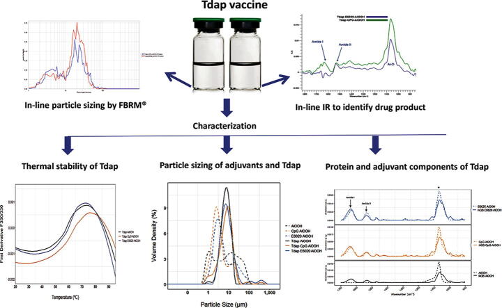
The goal of this study was to characterize an acellular pertussis vaccine (Tdap) containing genetically modified pertussis toxin (gdPT) and TLR agonist adsorbed to AlOOH adjuvant.
Several analytical tools including nanoDSF, FTIR, and LD were used to examine the conformation of novel gdPT and the composition of AlOOH adjuvant formulations adsorbed to pertussis vaccine.
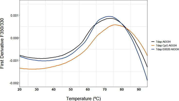
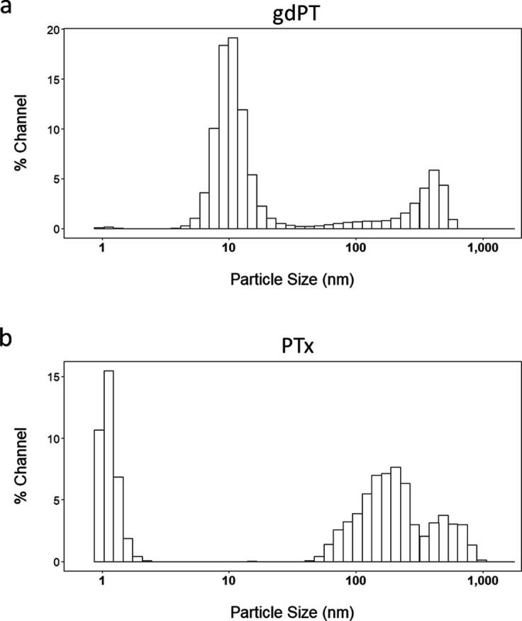
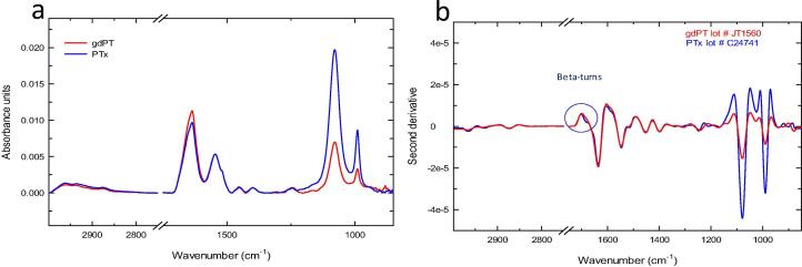
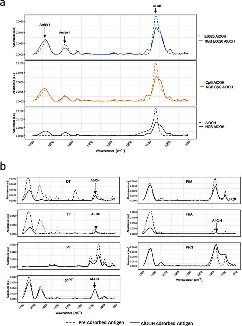
DLS particle size results were 9.3 nm and 320 nm for gdPT. For pertussis toxoid (PT), the DLS particle size results were larger at ~440 nm. After adsorption to AlOOH, which was driven by the protein antigen, the size distribution ranged from 3.5 to 22 µm. Two thermal transitions were observed by DSC for gdPT at 70 °C and 102 °C. The main thermal transition was confirmed to be at 72 °C by nanoDSF. All three vaccine formulations showed one thermal transition: Tdap-AlOOH had a thermal transition of 74.6 °C, Tdap-E6020-AlOOH had a thermal transition at 74.2 °C, and Tdap-CpG-AlOOH had a thermal transition at 77.0 °C. Analysis of pertussis toxin (PTx) and gdPT was also performed by FTIR spectroscopy for the purpose of comparison. The second derivative of the FTIR spectra showed an additional feature for PTx at 1685 cm compared to gdPT. The antigen's amide I and II regions were largely unchanged after adsorption to AlOOH adjuvant as shown by FTIR, suggesting that there were no significant changes in the secondary structure.
gdPT conformation was successfully characterized using an array of analytical methods. All three Tdap formulations have similar thermal stability as shown by nanoDSF, similar size distribution as shown by LD, and similar overall secondary structure as shown by FTIR. In-line particle sizing and IR can be used as in-process characterization tools to monitor consistency of adsorbed vaccine and to confirm product identity.
本研究的目的是对一种含有基因改造百日咳毒素(gdPT)和吸附于氢氧化铝佐剂的TLR激动剂的无细胞百日咳疫苗(Tdap)进行表征。
使用了包括纳米差示扫描荧光法(nanoDSF)、傅里叶变换红外光谱法(FTIR)和激光衍射法(LD)在内的多种分析工具,来检测新型gdPT的构象以及吸附于百日咳疫苗的氢氧化铝佐剂配方的组成。
gdPT的动态光散射(DLS)粒径结果为9.3纳米和320纳米。对于百日咳类毒素(PT),DLS粒径结果更大,约为440纳米。在由蛋白质抗原驱动吸附到氢氧化铝后,粒径分布范围为3.5至22微米。差示扫描量热法(DSC)观察到gdPT在70°C和102°C有两个热转变。纳米差示扫描荧光法确认主要热转变温度为72°C。所有三种疫苗配方均显示出一个热转变:Tdap - 氢氧化铝的热转变温度为74.6°C,Tdap - E6020 - 氢氧化铝的热转变温度为74.2°C,Tdap - CpG - 氢氧化铝的热转变温度为77.0°C。为了进行比较,还通过傅里叶变换红外光谱法对百日咳毒素(PTx)和gdPT进行了分析。与gdPT相比,傅里叶变换红外光谱的二阶导数显示PTx在1685厘米处有一个额外特征。傅里叶变换红外光谱显示,抗原的酰胺I和II区域在吸附到氢氧化铝佐剂后基本未变,表明二级结构没有显著变化。
使用一系列分析方法成功表征了gdPT的构象。如纳米差示扫描荧光法所示,所有三种Tdap配方具有相似的热稳定性;如激光衍射法所示,具有相似的粒径分布;如傅里叶变换红外光谱法所示,具有相似的整体二级结构。在线粒径分析和红外光谱可作为过程表征工具,以监测吸附疫苗的一致性并确认产品特性。