• 文献检索
  • 文档翻译
  • 深度研究
  • 学术资讯
  • Suppr Zotero 插件Zotero 插件
  • 邀请有礼
  • 套餐&价格
  • 历史记录
应用&插件
Suppr Zotero 插件Zotero 插件浏览器插件Mac 客户端Windows 客户端微信小程序
定价
高级版会员购买积分包购买API积分包
服务
文献检索文档翻译深度研究API 文档MCP 服务
关于我们
关于 Suppr公司介绍联系我们用户协议隐私条款
关注我们

Suppr 超能文献

核心技术专利:CN118964589B侵权必究
粤ICP备2023148730 号-1Suppr @ 2026

文献检索

告别复杂PubMed语法,用中文像聊天一样搜索,搜遍4000万医学文献。AI智能推荐,让科研检索更轻松。

立即免费搜索

文件翻译

保留排版,准确专业,支持PDF/Word/PPT等文件格式,支持 12+语言互译。

免费翻译文档

深度研究

AI帮你快速写综述,25分钟生成高质量综述,智能提取关键信息,辅助科研写作。

立即免费体验

通过透射电子显微镜正确识别冠状病毒的最佳实践。

Best practices for correctly identifying coronavirus by transmission electron microscopy.

机构信息

Synergy America, Inc., Atlanta, Georgia, USA.

Infectious Diseases Pathology Branch, Centers for Disease Control and Prevention (CDC), Atlanta, Georgia, USA.

出版信息

Kidney Int. 2021 Apr;99(4):824-827. doi: 10.1016/j.kint.2021.01.004. Epub 2021 Jan 22.

DOI:10.1016/j.kint.2021.01.004
PMID:33493525
原文链接:https://pmc.ncbi.nlm.nih.gov/articles/PMC7825881/
Abstract

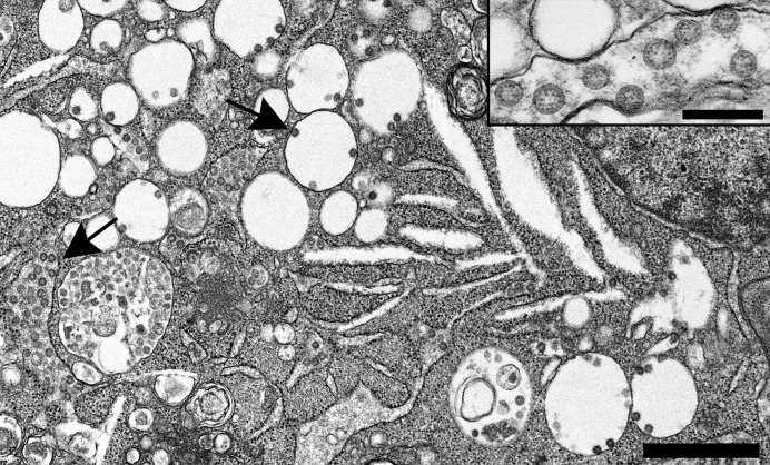
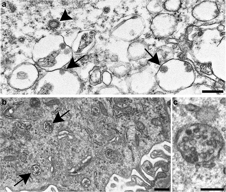
This guidance provides clear, concise strategies for identifying coronaviruses by transmission electron microscopy of ultrathin sections of tissues or infected tissue cultures. These include a description of virus morphology as well as cell organelles that can resemble viruses. Biochemical testing and caveats are discussed. Numerous references provide information for documentation and further study.

摘要

本指南提供了通过组织或感染组织培养物的超薄切片的透射电子显微镜来鉴定冠状病毒的清晰、简明的策略。其中包括对病毒形态以及可能类似于病毒的细胞细胞器的描述。还讨论了生化检测和注意事项。大量参考文献为文件记录和进一步研究提供了信息。

https://cdn.ncbi.nlm.nih.gov/pmc/blobs/f971/7825881/f57977c9b7f8/gr2_lrg.jpg
https://cdn.ncbi.nlm.nih.gov/pmc/blobs/f971/7825881/c85e02ce8827/gr1_lrg.jpg
https://cdn.ncbi.nlm.nih.gov/pmc/blobs/f971/7825881/f57977c9b7f8/gr2_lrg.jpg
https://cdn.ncbi.nlm.nih.gov/pmc/blobs/f971/7825881/c85e02ce8827/gr1_lrg.jpg
https://cdn.ncbi.nlm.nih.gov/pmc/blobs/f971/7825881/f57977c9b7f8/gr2_lrg.jpg

相似文献

1
Best practices for correctly identifying coronavirus by transmission electron microscopy.通过透射电子显微镜正确识别冠状病毒的最佳实践。
Kidney Int. 2021 Apr;99(4):824-827. doi: 10.1016/j.kint.2021.01.004. Epub 2021 Jan 22.
2
Hunting coronavirus by transmission electron microscopy - a guide to SARS-CoV-2-associated ultrastructural pathology in COVID-19 tissues.透射电镜检测冠状病毒——COVID-19 组织中 SARS-CoV-2 相关超微结构病理学的指南。
Histopathology. 2021 Feb;78(3):358-370. doi: 10.1111/his.14264. Epub 2020 Dec 1.
3
Difficulties in Differentiating Coronaviruses from Subcellular Structures in Human Tissues by Electron Microscopy.电子显微镜下鉴别人组织中冠状病毒与亚细胞结构的困难。
Emerg Infect Dis. 2021 Apr;27(4):1023-1031. doi: 10.3201/eid2704.204337. Epub 2021 Feb 18.
4
Severe acute respiratory syndrome coronavirus 2 (SARS-CoV2) culture and sample preparation for correlative light electron microscopy.严重急性呼吸综合征冠状病毒 2(SARS-CoV2)的培养和用于相关光电子显微镜术的样本准备。
Methods Cell Biol. 2024;187:99-116. doi: 10.1016/bs.mcb.2024.02.031. Epub 2024 Mar 4.
5
SARS-CoV-2: preliminary study of infected human nasopharyngeal tissue by high resolution microscopy.高分辨率显微镜观察感染 SARS-CoV-2 的人鼻咽组织的初步研究。
Virol J. 2021 Jul 18;18(1):149. doi: 10.1186/s12985-021-01620-1.
6
Comparison of RNA In Situ Hybridization and Immunohistochemistry Techniques for the Detection and Localization of SARS-CoV-2 in Human Tissues.RNA 原位杂交与免疫组织化学技术在检测和定位人组织中 SARS-CoV-2 的比较。
Am J Surg Pathol. 2021 Jan;45(1):14-24. doi: 10.1097/PAS.0000000000001563.
7
Detection and identification of coronaviruses in human tissues using electron microscopy.利用电子显微镜检测和识别人类组织中的冠状病毒。
Microsc Res Tech. 2022 Jul;85(7):2740-2747. doi: 10.1002/jemt.24115. Epub 2022 Apr 4.
8
Broad respiratory testing to identify SARS-CoV-2 viral co-circulation and inform diagnostic stewardship in the COVID-19 pandemic.广泛的呼吸检测以识别 SARS-CoV-2 病毒的共同传播并为 COVID-19 大流行中的诊断管理提供信息。
Virol J. 2021 May 1;18(1):93. doi: 10.1186/s12985-021-01545-9.
9
Just Seeing Is Not Enough for Believing: Immunolabelling as Indisputable Proof of SARS-CoV-2 Virions in Infected Tissue.眼见未必为实:免疫标记物无法作为新冠病毒感染组织中病毒粒子的无可争议证据。
Viruses. 2021 Sep 13;13(9):1816. doi: 10.3390/v13091816.
10
Ultrastructure of cell trafficking pathways and coronavirus: how to recognise the wolf amongst the sheep.细胞运输途径和冠状病毒的超微结构:如何在羊群中识别狼。
J Pathol. 2020 Dec;252(4):346-357. doi: 10.1002/path.5547. Epub 2020 Oct 22.

引用本文的文献

1
SARS-CoV-2 exploits steroidogenic machinery, triggers lipid metabolism for viral replication and induces immune response in Leydig cells of K18-hACE2 mice.严重急性呼吸综合征冠状病毒2(SARS-CoV-2)利用类固醇生成机制,触发脂质代谢以进行病毒复制,并在K18-hACE2小鼠的睾丸间质细胞中诱导免疫反应。
Front Cell Infect Microbiol. 2025 May 27;15:1538461. doi: 10.3389/fcimb.2025.1538461. eCollection 2025.
2
Study of histopathological and ultrastructural changes in the lungs of COVID-19 patients.新型冠状病毒肺炎患者肺部组织病理学及超微结构变化的研究
Med J Armed Forces India. 2025 May-Jun;81(3):328-336. doi: 10.1016/j.mjafi.2024.08.006. Epub 2024 Sep 19.
3

本文引用的文献

1
Appearances Can Be Deceiving - Viral-like Inclusions in COVID-19 Negative Renal Biopsies by Electron Microscopy.表象可能具有欺骗性——新冠病毒阴性肾活检组织电镜下类似病毒的包涵体
Kidney360. 2020 Jun 30;1(8):824-828. doi: 10.34067/KID.0002692020. eCollection 2020 Aug 27.
2
Difficulties in Differentiating Coronaviruses from Subcellular Structures in Human Tissues by Electron Microscopy.电子显微镜下鉴别人组织中冠状病毒与亚细胞结构的困难。
Emerg Infect Dis. 2021 Apr;27(4):1023-1031. doi: 10.3201/eid2704.204337. Epub 2021 Feb 18.
3
Why misinterpretation of electron micrographs in SARS-CoV-2-infected tissue goes viral.
Mouse hepatitis virus JHMV I protein is required for maximal virulence.
鼠肝炎病毒 JHMV I 蛋白是最大毒力所必需的。
J Virol. 2024 Sep 17;98(9):e0068024. doi: 10.1128/jvi.00680-24. Epub 2024 Aug 19.
4
Submandibular Gland Pathogenesis Following SARS-CoV-2 Infection and Implications for Xerostomia.感染 SARS-CoV-2 后下颌下腺的发病机制及其对口干症的影响。
Int J Mol Sci. 2024 Jun 21;25(13):6820. doi: 10.3390/ijms25136820.
5
Reconstruction of the real 3D shape of the SARS-CoV-2 virus.严重急性呼吸综合征冠状病毒2(SARS-CoV-2)病毒真实三维形状的重建。
Biophys J. 2024 May 21;123(10):1297-1310. doi: 10.1016/j.bpj.2024.04.019. Epub 2024 May 6.
6
Who Is the Intermediate Host of RNA Viruses? A Study Focusing on SARS-CoV-2 and Poliovirus.谁是RNA病毒的中间宿主?一项聚焦于新冠病毒和脊髓灰质炎病毒的研究。
Microorganisms. 2024 Mar 23;12(4):643. doi: 10.3390/microorganisms12040643.
7
Visualization of SARS-CoV-2 particles in naso/oropharyngeal swabs by thin section electron microscopy.通过超薄切片电子显微镜观察鼻咽拭子中的 SARS-CoV-2 病毒颗粒。
Virol J. 2023 Feb 6;20(1):21. doi: 10.1186/s12985-023-01981-9.
8
Ultrastructural Changes in Autopsy Tissues of COVID-19 Patients.新型冠状病毒肺炎患者尸检组织的超微结构变化
Cureus. 2022 Nov 27;14(11):e31932. doi: 10.7759/cureus.31932. eCollection 2022 Nov.
9
SARS-CoV-2: A Master of Immune Evasion.严重急性呼吸综合征冠状病毒2:免疫逃逸的高手
Biomedicines. 2022 Jun 7;10(6):1339. doi: 10.3390/biomedicines10061339.
10
SARS-CoV-2 Virology.SARS-CoV-2 病毒学。
Infect Dis Clin North Am. 2022 Jun;36(2):251-265. doi: 10.1016/j.idc.2022.01.004. Epub 2022 Jan 31.
为何新冠病毒感染组织中电子显微镜照片的错误解读会广泛传播。
Lancet. 2020 Oct 31;396(10260):e64-e65. doi: 10.1016/S0140-6736(20)32079-1. Epub 2020 Oct 5.
4
Hunting coronavirus by transmission electron microscopy - a guide to SARS-CoV-2-associated ultrastructural pathology in COVID-19 tissues.透射电镜检测冠状病毒——COVID-19 组织中 SARS-CoV-2 相关超微结构病理学的指南。
Histopathology. 2021 Feb;78(3):358-370. doi: 10.1111/his.14264. Epub 2020 Dec 1.
5
Postmortem Findings in Italian Patients With COVID-19: A Descriptive Full Autopsy Study of Cases With and Without Comorbidities.意大利 COVID-19 患者的尸检结果:有和无合并症病例的描述性全面尸检研究。
J Infect Dis. 2020 Nov 9;222(11):1807-1815. doi: 10.1093/infdis/jiaa578.
6
SARS-CoV-2 causes a specific dysfunction of the kidney proximal tubule.SARS-CoV-2 导致肾脏近端小管的特定功能障碍。
Kidney Int. 2020 Nov;98(5):1296-1307. doi: 10.1016/j.kint.2020.07.019. Epub 2020 Aug 10.
7
Fatal Pulmonary Thromboembolism in SARS-CoV-2-Infection.新型冠状病毒感染导致的致命性肺血栓栓塞症
Cardiovasc Pathol. 2020 Sep-Oct;48:107227. doi: 10.1016/j.carpath.2020.107227. Epub 2020 May 12.
8
Histopathology and ultrastructural findings of fatal COVID-19 infections in Washington State: a case series.华盛顿州致命性 COVID-19 感染的组织病理学和超微结构发现:病例系列研究。
Lancet. 2020 Aug 1;396(10247):320-332. doi: 10.1016/S0140-6736(20)31305-2. Epub 2020 Jul 16.
9
Pulmonary Vascular Pathology in Covid-19. Reply.新型冠状病毒肺炎中的肺血管病理学。回复
N Engl J Med. 2020 Aug 27;383(9):888-889. doi: 10.1056/NEJMc2022068. Epub 2020 Jul 17.
10
Caution in Identifying Coronaviruses by Electron Microscopy.通过电子显微镜鉴定冠状病毒时需谨慎。
J Am Soc Nephrol. 2020 Sep;31(9):2223-2224. doi: 10.1681/ASN.2020050755. Epub 2020 Jul 10.