Department of Biomedical Engineering, University of California-Irvine, Irvine, CA, USA.
Department of Anatomy and Physiology, Queens College, City University of New York, Bayside, NY, USA.
Sci Rep. 2021 Jan 25;11(1):2141. doi: 10.1038/s41598-021-81534-8.
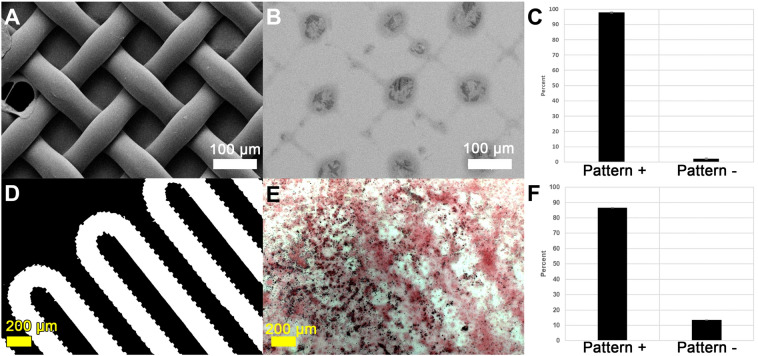
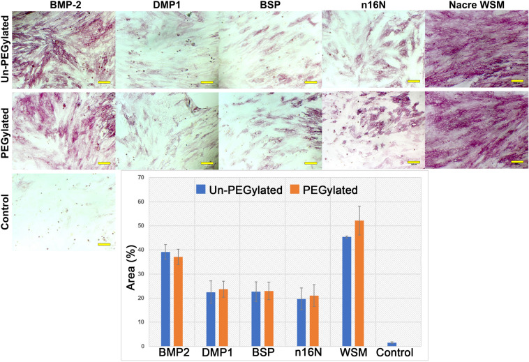
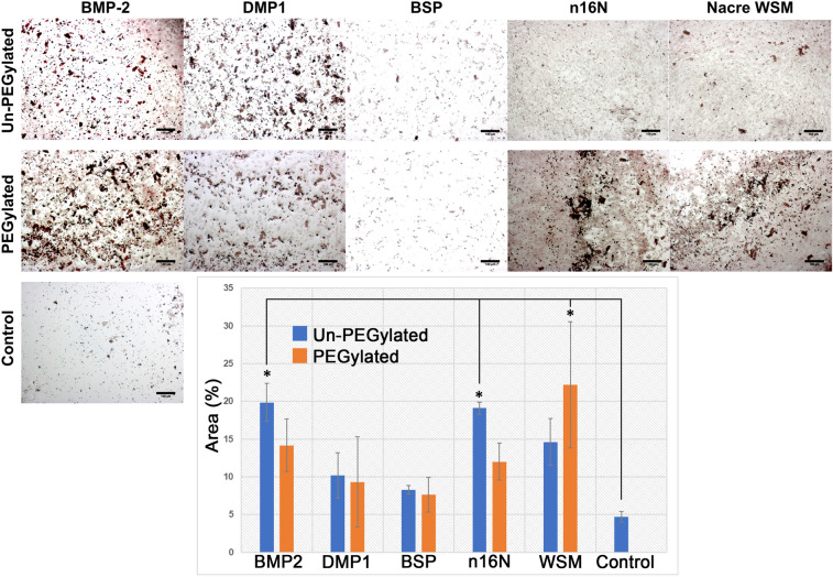
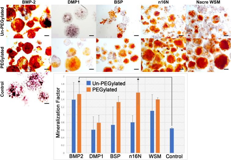
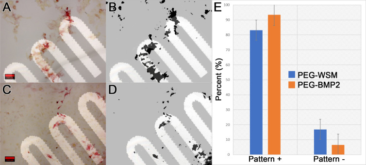
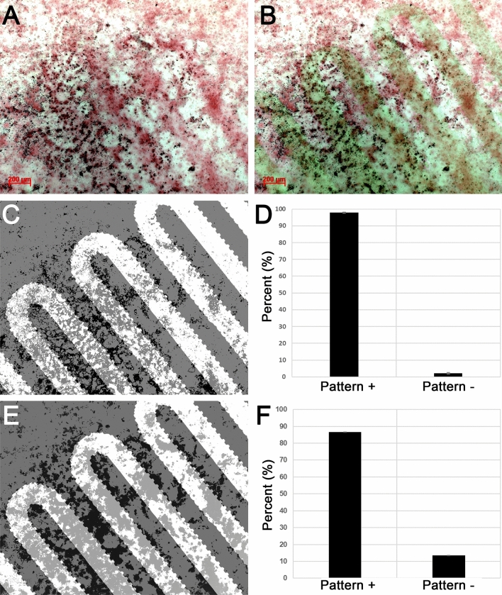
In response to the drawbacks of autograft donor-site morbidity and bone morphogenetic protein type 2 (BMP2) carcinogenesis and ectopic bone formation, there has been an increased research focus towards developing alternatives capable of achieving spatial control over bone formation. Here we show for the first time both osteogenic differentiation and mineralization (from solution or mediated by cells) occurring within predetermined microscopic patterns. Our results revealed that both PEGylated BMP2 and nacre proteins induced stem cell osteodifferentiation in microscopic patterns when these proteins were covalently bonded in patterns onto polyethylene glycol diacrylate (PEGDA) hydrogel substrates; however, only nacre proteins induced mineralization localized to the micropatterns. These findings have broad implications on the design and development of orthopedic biomaterials and drug delivery.
为了解决自体移植物供体部位发病率和骨形态发生蛋白 2(BMP2)致癌和异位骨形成的缺点,人们越来越关注开发能够实现对骨形成的空间控制的替代物。在这里,我们首次展示了在预定的微观图案内发生的成骨分化和矿化(来自溶液或通过细胞介导)。我们的结果表明,当这些蛋白质在图案中键合到聚乙二醇二丙烯酸酯(PEGDA)水凝胶基底上时,PEGylated BMP2 和珍珠层蛋白都可以诱导干细胞在微观图案中发生成骨分化;但是,只有珍珠层蛋白可以诱导矿化定位于微图案。这些发现对骨科生物材料和药物输送的设计和开发具有广泛的意义。