Department of Radiology and Imaging, Hospital for Special Surgery, New York, New York, USA.
Departments of Physiatry and Sports Medicine, Hospital for Special Surgery, New York, New York, USA.
Muscle Nerve. 2021 May;63(5):703-709. doi: 10.1002/mus.27186. Epub 2021 Feb 12.
In this study, we aimed to determine whether muscle transverse relaxation time (T ) magnetic resonance (MR) mapping results correlate with motor unit loss, as defined by motor unit recruitment patterns on electromyography (EMG).
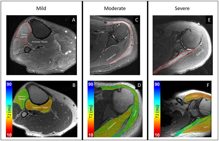
EMG and 3-Tesla MRI exams were acquired no more than 31 days apart in subjects referred for peripheral nerve MRI. Two musculoskeletal radiologists qualitatively graded T -weighted, fat-suppressed sequences for severity of muscle edema-like patterns and manually placed regions of interest within muscles to obtain T values from T -mapping sequences. Concordance was calculated between qualitative and quantitative MR grades and EMG recruitment categories (none, discrete, decreased) as well as interobserver agreement for both MR grades.
Thirty-four muscles (21 abnormal, 13 control) were assessed in 13 subjects (5 females and 8 males; mean age, 46 years) with 14 EMG-MRI pairs. T -relaxation times were significantly (P < .001) increased in all EMG recruitment categories compared with control muscles. T differences were not significant between EMG grades of motor unit recruitment (P = .151-.702). T and EMG score concordance was acceptable (Harrell's concordance index [c index]: rater A, 0.71; 95% confidence interval [CI], 0.51-0.87; rater B, 0.77; 95% CI, 0.57-0.91). Qualitative MRI and EMG score concordance was poor to acceptable (c index: rater A, 0.60; 95% CI, 0.50-0.79; rater B, 0.72; 95% CI, 0.55-0.89). T values had moderate-to-substantial ability to distinguish between absent vs incomplete (ie, decreased or discrete) motor unit recruitment (c index: rater A, 0.78; 95% CI, 0.50-1.00; rater B, 0.86; 95% CI, 0.57-1.00).
Quantitative T MR muscle mapping is a promising tool for noninvasive evaluation of the degree of motor unit recruitment loss.
本研究旨在确定磁共振(MR)T2 弛豫时间(T2)映射结果是否与肌电图(EMG)定义的运动单位丧失相关。
在进行外周神经 MRI 检查的患者中,EMG 和 3T MRI 检查的时间间隔不超过 31 天。两名肌肉骨骼放射科医生对 T2 加权、脂肪抑制序列进行定性评估,以评估肌肉水肿样模式的严重程度,并手动在肌肉内放置感兴趣区,以从 T 映射序列中获取 T 值。评估了 13 名患者(5 名女性和 8 名男性;平均年龄 46 岁)的 34 块肌肉(21 块异常,13 块正常),其中有 14 对 EMG-MRI。与正常肌肉相比,所有 EMG 募集类别(离散募集、递减募集、无募集)的 T 弛豫时间均显著增加(P < 0.001)。EMG 募集程度之间的 T 差异无统计学意义(P = 0.151-0.702)。T 值与 EMG 评分的一致性尚可(哈雷尔一致性指数[c 指数]:A 评分者为 0.71;95%置信区间[CI]为 0.51-0.87;B 评分者为 0.77;95%CI 为 0.57-0.91)。T 值和 EMG 评分的一致性从差到尚可(c 指数:A 评分者为 0.60;95%CI 为 0.50-0.79;B 评分者为 0.72;95%CI 为 0.55-0.89)。T 值能够很好地区分缺失与不完全(即递减或离散)运动单位募集(c 指数:A 评分者为 0.78;95%CI 为 0.50-1.00;B 评分者为 0.86;95%CI 为 0.57-1.00)。
定量 T2 弛豫时间磁共振肌肉成像可能是一种有前途的非侵入性工具,用于评估运动单位募集丧失的程度。