Department of Nuclear Medicine, University of Duisburg-Essen and German Cancer Consortium-University Hospital Essen, Essen, Germany.
Department of Gastroenterology and Hepatology, University Hospital Essen, University of Duisburg-Essen, Essen, Germany; and.
J Nucl Med. 2021 Sep 1;62(9):1235-1241. doi: 10.2967/jnumed.120.257915. Epub 2021 Jan 28.
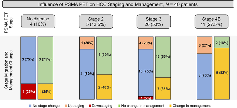
Hepatocellular carcinoma (HCC) is the sixth most prevalent cancer and the third most frequent cause of cancer-related death. A growing number of local and systemic therapies are available, and accurate staging is critical for management decisions. We assessed the impact of neovasculature imaging by Ga-PSMA-11 PET/CT on disease staging, prognostic groups, and management of patients with HCC compared with staging with CT. Forty patients who received imaging with Ga-PSMA-11 PET/CT for HCC staging between September 2018 and September 2019 were retrospectively included. Management before and after PET scanning was assessed by standardized surveys. The presence of HCC was evaluated by 3 masked readers on a per-patient and per-region basis for PET/CT (PET criteria) and multiphase contrast-enhanced CT (CT criteria) in separate sessions. Lesions were validated by follow-up imaging or histopathology, and progression-free survival was recorded. Endpoints were detection rate and positive predictive value for Ga-PSMA-11 PET versus CT, interreader reproducibility, and changes in stage, prognostic groups, and management plans. Median age was 65 y (range, 37-81 y), and median Child-Pugh score was 5 (range, 5-9). Most patients were treatment-naïve (27/40, 67.5%). The sensitivity of PET versus CT to identify liver lesions for patients with lesion validation was 31 of 32 (97%) for both modalities, whereas it was 6 of 6 (100%) versus 4 of 6 (67%), respectively, for extrahepatic lesions. PET and CT each had a positive predictive value of 100% at the liver level. PET versus CT stage was congruent in 30 of 40 (75%) patients; upstaging was seen in 8 of 40 patients (20%), whereas 2 of 40 (5%) had downstaging by PET. Intended management changed in 19 of 40 patients (47.5%); 9 of 19 of these patients were found to have detectable distant metastases (47.4%) and assigned stage 4 disease, most of whom were shifted to systemic therapy (8/9, 89%). Two patients underwent Lu-PSMA-617 radioligand therapy. Median progression-free survival was 5.2 mo for the entire cohort; 5.3 mo for PET M0, and 4.7 mo for PET M1 patients, respectively. Ga-PSMA-11 PET demonstrated higher accuracy than CT in the detection of HCC metastases and was associated with a management change in about half the patient cohort.
肝细胞癌(HCC)是第六大常见癌症,也是癌症相关死亡的第三大常见原因。越来越多的局部和全身治疗方法可供选择,准确分期对于管理决策至关重要。我们评估了 Ga-PSMA-11 PET/CT 对 HCC 分期、预后分组和治疗的影响,与 CT 相比。
回顾性纳入了 2018 年 9 月至 2019 年 9 月间因 HCC 分期接受 Ga-PSMA-11 PET/CT 成像的 40 例患者。通过标准化调查评估 PET 扫描前后的管理情况。对每位患者和每个区域的 PET/CT(PET 标准)和多相增强 CT(CT 标准)进行了 3 位盲法读者的评估。在单独的会议上,通过随访成像或组织病理学对病变进行验证,并记录无进展生存期。终点为 Ga-PSMA-11 PET 与 CT 的检测率和阳性预测值、读者间的可重复性以及分期、预后分组和管理计划的变化。
中位年龄为 65 岁(范围 37-81 岁),中位 Child-Pugh 评分为 5 分(范围 5-9 分)。大多数患者为初治(27/40,67.5%)。对于有病变验证的患者,PET 与 CT 对肝脏病变的检出率分别为 31/32(97%)和 6/6(100%),而对于肝外病变,分别为 4/6(67%)和 6/6(100%)。PET 和 CT 在肝脏水平的阳性预测值均为 100%。40 例患者中,PET 与 CT 分期一致的有 30 例(75%);40 例患者中有 8 例(20%)分期升高,2 例(5%)PET 分期降低。40 例患者中有 19 例(47.5%)的治疗计划发生改变;19 例中有 9 例(47.4%)患者发现有可检测的远处转移,并被归为 4 期疾病,其中大多数患者被转为全身治疗(8/9,89%)。2 例患者接受了 Lu-PSMA-617 放射性配体治疗。整个队列的中位无进展生存期为 5.2 个月;PET M0 为 5.3 个月,PET M1 患者为 4.7 个月。与 CT 相比,Ga-PSMA-11 PET 在 HCC 转移的检测中具有更高的准确性,并与约一半的患者队列的治疗计划改变相关。