Sun Xingwei, Zhou Feng, Bai Xuming, Yuan Qiang, Zhang Mingqing, Ma Liang, Jin Yong
Department of Intervention, The Second Affiliated Hospital of Soochow University, No.1055 Sanxiang Road, Suzhou, Jiangsu, 215004, People's Republic of China.
Department of Ultrasound Medicine, The Affiliated Suzhou Science & Technology Town Hospital of Nanjing Medical University, Suzhou, 215000, People's Republic of China.
World J Surg Oncol. 2021 Jan 30;19(1):32. doi: 10.1186/s12957-021-02144-2.
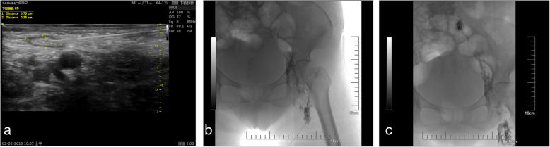
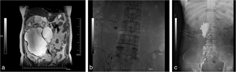
Traumatic lymphatic leakage is a rare but potentially life-threatening complication. The purpose of this study was to introduce ultrasound-guided intranodal lymphangiography and embolisation techniques for postoperative lymphatic leakage in patients with cancer.
From January 2018 through June 2020, seven cancer patients (three males, four females, aged 59-75 years [mean 67.57 ± 6.11 years]) developed lymphatic leakage after abdominal or pelvic surgery, with drainage volumes ranging from 550 to 1200 mL per day. The procedure and follow-up of ultrasound-guided intranodal lymphangiography and embolisation were recorded. This study retrospectively analysed the technical success rate, operative time, length of hospital stay, clinical efficacy, and complications.
The operation was technically successful in all patients. Angiography revealed leakage, and embolisation was performed in all seven patients (7/7, 100%). The operative time of angiography and embolisation was 41 to 68 min, with an average time of 53.29 ± 10.27 min. The mean length of stay was 3.51 ± 1.13 days. Lymph node embolisation was clinically successful in five patients (5/7, 71.43%), who had a significant reduction in or disappearance of chylous ascites. The other two patients received surgical treatment 2 weeks later due to poor results after embolisation. All patients were followed for 2 weeks. No serious complications or only minor complications were found in all the patients.
Ultrasound-guided intranodal lymphangiography and embolisation were well tolerated by the patients, who experienced a low incidence of complications. Early intervention is recommended for cancer patients with postoperative lymphatic leakage.
创伤性淋巴漏是一种罕见但可能危及生命的并发症。本研究的目的是介绍超声引导下淋巴结内淋巴管造影及栓塞技术用于癌症患者术后淋巴漏的治疗。
2018年1月至2020年6月,7例癌症患者(3例男性,4例女性,年龄59 - 75岁[平均67.57±6.11岁])在腹部或盆腔手术后发生淋巴漏,每日引流量为550至1200毫升。记录超声引导下淋巴结内淋巴管造影及栓塞的操作过程和随访情况。本研究回顾性分析了技术成功率、手术时间、住院时间、临床疗效及并发症。
所有患者手术技术均成功。血管造影显示有渗漏,7例患者均进行了栓塞(7/7,100%)。血管造影及栓塞的手术时间为41至68分钟,平均时间为53.29±10.27分钟。平均住院时间为3.51±1.13天。5例患者(5/7,71.43%)淋巴结栓塞临床成功,乳糜腹水明显减少或消失。另外2例患者因栓塞效果不佳在2周后接受了手术治疗。所有患者均随访2周。所有患者均未发现严重并发症或仅出现轻微并发症。
患者对超声引导下淋巴结内淋巴管造影及栓塞耐受性良好,并发症发生率低。建议对癌症术后淋巴漏患者进行早期干预。