Department of Oral and Cranio-Maxillofacial Surgery, Ninth People's Hospital, Shanghai Jiao Tong University School of Medicine, Shanghai, 200011, China.
National Clinical Research Center for Oral Diseases, Shanghai, 200011, China.
Orphanet J Rare Dis. 2021 Jan 30;16(1):59. doi: 10.1186/s13023-021-01713-8.
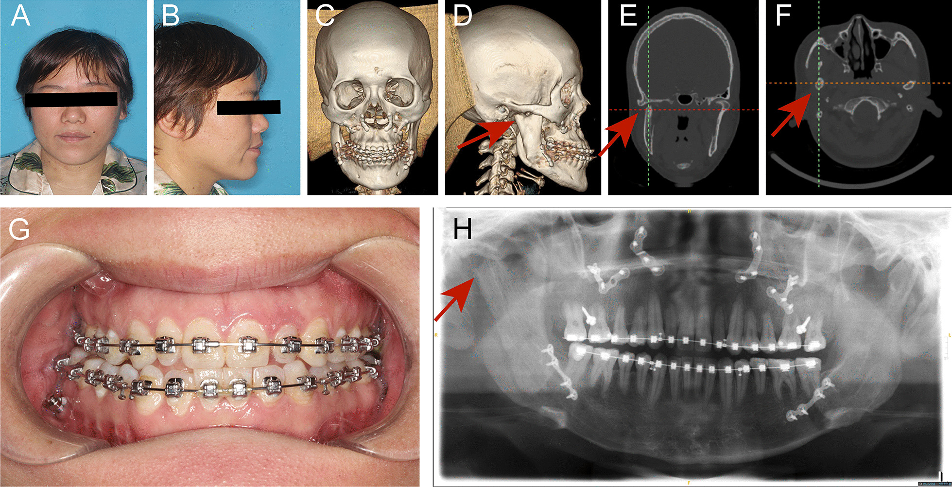
Mandibular condylar osteochondroma (OC) could lead to facial morphologic and functional disturbances, such as facial asymmetry, malocclusion, and temporomandibular joint dysfunction. However, after condylar OC resection, the inaccurate reposition of the neocondyle still needs to be solved. The purpose of this study was to explore the feasibility of the condylar osteotomy and repositioning guide to reposition the neocondyle in the treatment of patients with severe deformity secondary to condylar OC.
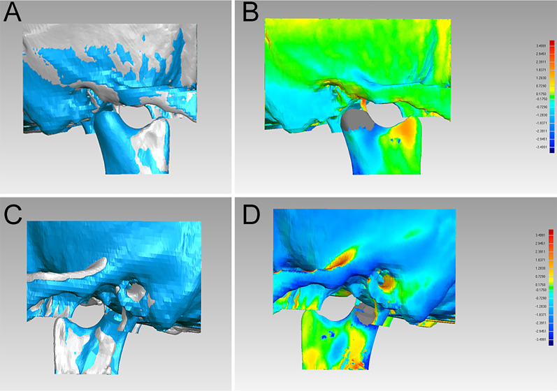
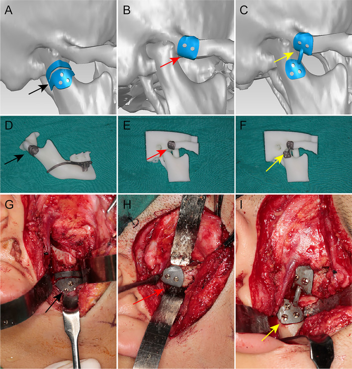
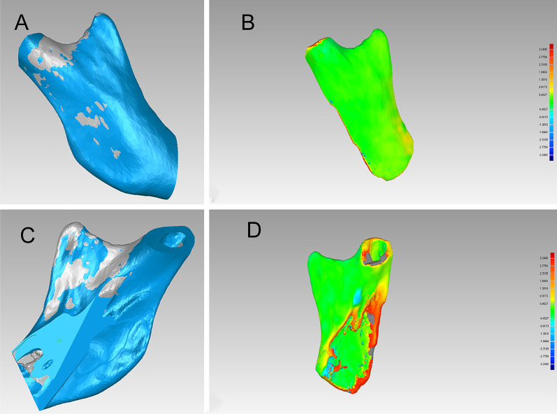
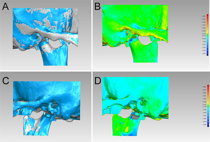
Three patients with severe deformity secondary to OC of the mandibular condyle were enrolled in this study. With the aid of condylar osteotomy and repositioning guide, condylar OC resection and repositioning were carried out, and the accuracy and stability of these guides were evaluated. All patients healed uneventfully, and no facial nerve injury and condylar ankylosis occurred. Compared with the computerized tomography scans in centric relation before surgery and 3 days after surgery, the results showed that the facial symmetry was greatly improved in all the patients. Also, after the superimposition of the condylar segments before surgery and 3 days after surgery, the postoperative reconstructed condyles had a high degree of similarity to the reconstruction of the virtual surgical planning. Observed from the sagittal and coronal directions, the measurements of condylar positions were very close to those of virtual surgical planning. Moreover, it also showed stable results after a 1-year follow-up.
For patients with severe deformity secondary to condylar OC, condylar osteotomy, and repositioning guide was expected to provide a new option for the improvement of facial symmetry and occlusal relationship.
下颌骨髁突骨软骨瘤(OC)可导致面部形态和功能紊乱,如面部不对称、咬合不正和颞下颌关节功能障碍。然而,在髁突 OC 切除后,新髁突的位置不准确仍需解决。本研究旨在探讨髁突切开复位导板在治疗严重畸形继发于髁突 OC 患者中的可行性。
本研究纳入了 3 名严重畸形继发于下颌骨髁突 OC 的患者。在髁突切开复位导板的辅助下,进行了髁突 OC 切除和复位,评估了这些导板的准确性和稳定性。所有患者均顺利愈合,无面神经损伤和髁突强直发生。与术前和术后 3 天正中关系位的计算机断层扫描相比,所有患者的面部对称性均得到了显著改善。此外,在术前和术后 3 天的髁突节段叠加后,术后重建的髁突与虚拟手术规划的重建具有高度相似性。从矢状面和冠状面观察,髁突位置的测量值非常接近虚拟手术规划。而且,1 年随访后也显示出稳定的结果。
对于严重畸形继发于髁突 OC 的患者,髁突切开复位导板有望为改善面部对称性和咬合关系提供新的选择。