• 文献检索
  • 文档翻译
  • 深度研究
  • 学术资讯
  • Suppr Zotero 插件Zotero 插件
  • 邀请有礼
  • 套餐&价格
  • 历史记录
应用&插件
Suppr Zotero 插件Zotero 插件浏览器插件Mac 客户端Windows 客户端微信小程序
定价
高级版会员购买积分包购买API积分包
服务
文献检索文档翻译深度研究API 文档MCP 服务
关于我们
关于 Suppr公司介绍联系我们用户协议隐私条款
关注我们

Suppr 超能文献

核心技术专利:CN118964589B侵权必究
粤ICP备2023148730 号-1Suppr @ 2026

文献检索

告别复杂PubMed语法,用中文像聊天一样搜索,搜遍4000万医学文献。AI智能推荐,让科研检索更轻松。

立即免费搜索

文件翻译

保留排版,准确专业,支持PDF/Word/PPT等文件格式,支持 12+语言互译。

免费翻译文档

深度研究

AI帮你快速写综述,25分钟生成高质量综述,智能提取关键信息,辅助科研写作。

立即免费体验

计算机辅助双侧矢状劈开截骨术教学:髁突定位的学习曲线。

Computer-assisted teaching of bilateral sagittal split osteotomy: Learning curve for condylar positioning.

机构信息

Department of Oral and Maxillofacial Surgery, Head and Neck Institute, University Hospital of Nice, Nice, France.

Biostatistic Department, Centre Antoine Lacassagne, Nice, France.

出版信息

PLoS One. 2018 Apr 25;13(4):e0196136. doi: 10.1371/journal.pone.0196136. eCollection 2018.

DOI:10.1371/journal.pone.0196136
PMID:29694423
原文链接:https://pmc.ncbi.nlm.nih.gov/articles/PMC5918964/
Abstract

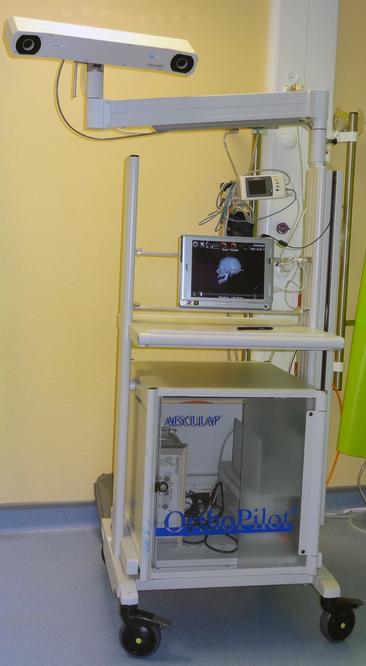
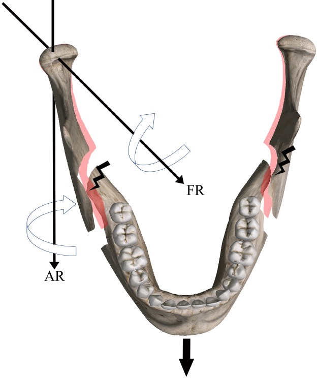
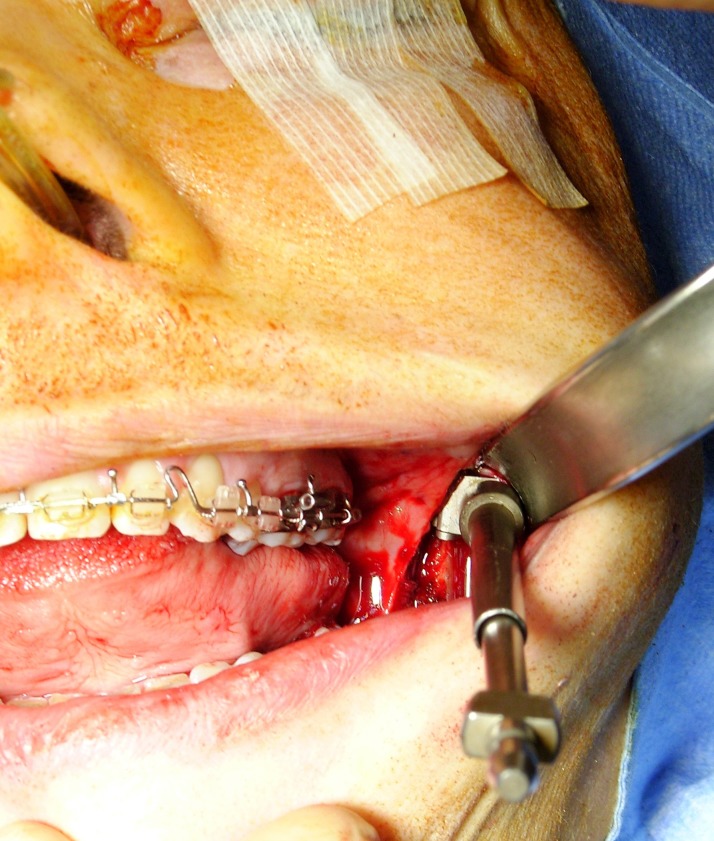
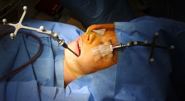
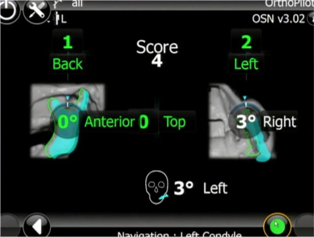
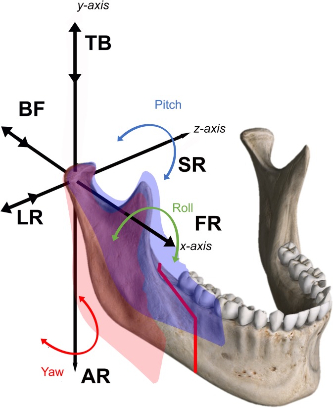
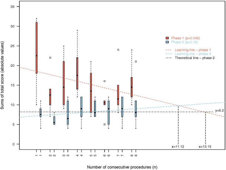
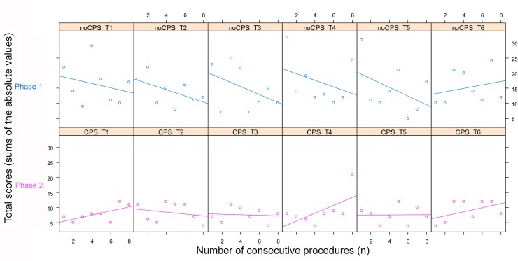
Bilateral sagittal split osteotomy (BSSO) is a widely-performed procedure in orthognathic surgery for the correction of dentofacial deformity. Condylar positioning is a critical step during BSSO to maximize functional and morphological results. The unsuitable positioning of condyles represents one of the causative mechanisms that may induce temporomandibular joint noxious effects after BSSO. Repositioning devices can assist surgeons in maintaining the preoperative condylar position; however, empirical repositioning methods based on experience gained are still commonly used. Trainee learning curves are difficult to assess. The aim of this study was to evaluate the relevance of computer-assisted surgery in the acquisition of condylar positioning skills. Forty-eight patients underwent BSSO performed by six maxillofacial trainees (four junior residents and two senior experienced residents). A condyle positioning system (CPS) was used by a senior surgeon to record a condylar position score during the procedure. Firstly, scores were recorded when the trainee manually positioned the condyle without access to the CPS score (phase 1) and then when the trainee positioned the condyle and performed osteosynthesis with visual access to the CPS score (phase 2). Six parameters describing condylar three-dimensional motions were assessed: translational motion from top to bottom (TB), back to front (BF), and left to right (LR), axial rotation (AR), sagittal rotation (SR), frontal rotation (FR), and a total score (TS). There were no significant differences between junior and senior residents in condyle positioning without access to the CPS. Condyles were significantly better positioned during phase 2 with access to the CPS (p<0.001). Over time, use of the CPS (phase 2) produced significantly quicker improvements in scores (p = 0.042). For those teaching surgeries to trainees, computer-assisted devices can potentially result in more rapid learning curves than traditional "observations-imitation" models. Use of a CPS by trainees facilitated condylar repositioning that resulted in an accurate occlusal result and avoidance of adverse effects on the temporomandibular joint.

摘要

双侧矢状劈开截骨术(Bilateral Sagittal Split Osteotomy,BSSO)是正颌外科中用于矫正牙颌面畸形的广泛应用的手术方法。髁突定位是 BSSO 中至关重要的一步,旨在实现最佳的功能和形态效果。髁突位置不当是 BSSO 后可能引起颞下颌关节不良影响的一个致病机制。复位设备可以帮助外科医生维持术前髁突位置;然而,仍广泛采用基于经验的经验性复位方法。学员学习曲线难以评估。本研究旨在评估计算机辅助手术在获得髁突定位技能中的相关性。48 例患者接受了由六名颌面外科住院医师(四名初级住院医师和两名高级经验丰富的住院医师)进行的 BSSO。一名资深外科医生使用髁突定位系统(CPS)在手术过程中记录髁突位置评分。首先,在学员无法访问 CPS 评分的情况下手动定位髁突时记录评分(第 1 阶段),然后在学员定位髁突并在可视访问 CPS 评分的情况下进行骨合成时记录评分(第 2 阶段)。评估了描述髁突三维运动的六个参数:从上到下的平移运动(TB)、从后到前的平移运动(BF)和从左到右的平移运动(LR)、轴向旋转(AR)、矢状旋转(SR)、额状旋转(FR)和总得分(TS)。在无法访问 CPS 的情况下,初级和高级住院医师的髁突定位没有显著差异。在可视访问 CPS 时(第 2 阶段),髁突定位明显更好(p<0.001)。随着时间的推移,使用 CPS(第 2 阶段)可显著提高评分(p = 0.042)。对于那些向学员教授手术的人来说,计算机辅助设备可能会导致比传统的“观察-模仿”模型更快的学习曲线。学员使用 CPS 有助于髁突重新定位,从而获得准确的咬合结果,并避免对颞下颌关节产生不良影响。

https://cdn.ncbi.nlm.nih.gov/pmc/blobs/0d54/5918964/2f74ae4bbd6d/pone.0196136.g008.jpg
https://cdn.ncbi.nlm.nih.gov/pmc/blobs/0d54/5918964/82ccb6833bc9/pone.0196136.g001.jpg
https://cdn.ncbi.nlm.nih.gov/pmc/blobs/0d54/5918964/ea9f61a3eab5/pone.0196136.g002.jpg
https://cdn.ncbi.nlm.nih.gov/pmc/blobs/0d54/5918964/30e87cf8eee9/pone.0196136.g003.jpg
https://cdn.ncbi.nlm.nih.gov/pmc/blobs/0d54/5918964/66bea74f277d/pone.0196136.g004.jpg
https://cdn.ncbi.nlm.nih.gov/pmc/blobs/0d54/5918964/9ef4660d271b/pone.0196136.g005.jpg
https://cdn.ncbi.nlm.nih.gov/pmc/blobs/0d54/5918964/1fd1ab6501ba/pone.0196136.g006.jpg
https://cdn.ncbi.nlm.nih.gov/pmc/blobs/0d54/5918964/92e7d675dec0/pone.0196136.g007.jpg
https://cdn.ncbi.nlm.nih.gov/pmc/blobs/0d54/5918964/2f74ae4bbd6d/pone.0196136.g008.jpg

相似文献

1
Computer-assisted teaching of bilateral sagittal split osteotomy: Learning curve for condylar positioning.计算机辅助双侧矢状劈开截骨术教学:髁突定位的学习曲线。
PLoS One. 2018 Apr 25;13(4):e0196136. doi: 10.1371/journal.pone.0196136. eCollection 2018.
2
Accuracy of computer-assisted design and manufactured three-dimensional device for condylar positioning in mandibular bilateral sagittal split osteotomy (clinical trial).计算机辅助设计和制作的三维下颌骨双侧矢状劈开截骨术下颌髁突定位装置的准确性(临床试验)。
Oral Surg Oral Med Oral Pathol Oral Radiol. 2024 Apr;137(4):331-337. doi: 10.1016/j.oooo.2023.11.001. Epub 2023 Nov 8.
3
Evaluation of three-dimensional position change of the condylar head after orthognathic surgery using computer-aided design/computer-aided manufacturing-made condyle positioning jig.使用计算机辅助设计/计算机辅助制造的髁突定位夹具评估正颌外科手术后髁突头部的三维位置变化。
J Craniofac Surg. 2014 Nov;25(6):2002-7. doi: 10.1097/SCS.0b013e3182a246f1.
4
[Mandibular condyle localization in orthognathic surgery based on mandibular movement trajectory and its preliminary accuracy verification].基于下颌运动轨迹的正颌外科下颌髁突定位及其初步准确性验证
Beijing Da Xue Xue Bao Yi Xue Ban. 2024 Feb 18;56(1):57-65. doi: 10.19723/j.issn.1671-167X.2024.01.010.
5
Changes in Condylar Position Within 12 Months After Bilateral Sagittal Split Ramus Osteotomy With and Without Le Fort I Osteotomy by Using Cone-Beam Computed Tomography.利用锥形束计算机断层扫描评估双侧下颌升支矢状劈开截骨术联合或不联合 Le Fort I 截骨术后 12 个月髁突位置的变化。
J Oral Maxillofac Surg. 2022 Jan;80(1):162-173. doi: 10.1016/j.joms.2021.08.146. Epub 2021 Aug 17.
6
Changes of temporomandibular joint position after surgery first orthognathic treatment concept.手术后颞下颌关节位置的变化:正颌治疗理念的首次改变。
Sci Rep. 2019 Feb 18;9(1):2206. doi: 10.1038/s41598-019-38786-2.
7
A Modified Novel Technique for Condylar Positioning in Mandibular Bilateral Sagittal Split Osteotomy Using Computer-Assisted Designed and Computer-Assisted Manufactured Surgical Guides.一种使用计算机辅助设计和计算机辅助制造手术导板在下颌双侧矢状劈开截骨术中髁突定位的改良新技术。
J Oral Maxillofac Surg. 2019 May;77(5):1069.e1-1069.e9. doi: 10.1016/j.joms.2019.01.014.
8
Short- and long-term changes of condylar position after bilateral sagittal split ramus osteotomy for mandibular advancement in combination with Le Fort I osteotomy evaluated by cone-beam computed tomography.通过锥形束计算机断层扫描评估下颌前徙联合Le Fort I截骨术的双侧矢状劈开下颌支截骨术后髁突位置的短期和长期变化。
J Oral Maxillofac Surg. 2013 Nov;71(11):1956-66. doi: 10.1016/j.joms.2013.06.213. Epub 2013 Aug 22.
9
Does orthognathic surgery affect mandibular condyle position? A retrospective study.正颌手术是否会影响下颌髁突位置?一项回顾性研究。
Oral Maxillofac Surg. 2024 Jun;28(2):639-643. doi: 10.1007/s10006-023-01181-3. Epub 2023 Sep 23.
10
Intraoperative condyle positioning by sonographic monitoring in orthognathic surgery verified by MRI.正颌手术中通过超声监测髁突定位并经磁共振成像验证
J Craniomaxillofac Surg. 2015 Jan;43(1):71-80. doi: 10.1016/j.jcms.2014.10.012. Epub 2014 Oct 22.

引用本文的文献

1
Evaluation of mandibular condyle position in Class III patients after bimaxillary orthognathic surgery: A cone-beam computed tomography study.双颌正颌手术后III类患者下颌髁突位置的评估:一项锥形束计算机断层扫描研究。
Korean J Orthod. 2024 Jul 25;54(4):247-256. doi: 10.4041/kjod23.188. Epub 2024 Jul 10.
2
Comparison of Patient-Specific Condylar Positioning Devices and Manual Methods in Orthognathic Surgery: A Prospective Randomized Trial.正颌外科中患者特异性髁突定位装置与手动方法的比较:一项前瞻性随机试验
J Clin Med. 2024 Jan 27;13(3):737. doi: 10.3390/jcm13030737.
3
A new method for individual condylar osteotomy and repositioning guides used in patients with severe deformity secondary to condylar osteochondroma.

本文引用的文献

1
The use of patient-specific implants in orthognathic surgery: A series of 30 mandible sagittal split osteotomy patients.个性化植入物在正颌外科手术中的应用:30例下颌矢状劈开截骨术患者的系列研究。
J Craniomaxillofac Surg. 2017 Jun;45(6):990-994. doi: 10.1016/j.jcms.2017.02.021. Epub 2017 Mar 3.
2
Surgical Navigation: A Systematic Review of Indications, Treatments, and Outcomes in Oral and Maxillofacial Surgery.手术导航:口腔颌面外科手术适应症、治疗方法及结果的系统评价
J Oral Maxillofac Surg. 2017 Sep;75(9):1987-2005. doi: 10.1016/j.joms.2017.01.004. Epub 2017 Jan 18.
3
A review of computer-aided oral and maxillofacial surgery: planning, simulation and navigation.
一种用于因髁突骨软骨瘤导致严重畸形的患者的个体化髁突截骨和复位导板的新方法。
Orphanet J Rare Dis. 2021 Jan 30;16(1):59. doi: 10.1186/s13023-021-01713-8.
4
Fixation Methods for Mandibular Advancement and Their Effects on Temporomandibular Joint: A Finite Element Analysis Study.下颌前伸固定方法及其对颞下颌关节的影响的有限元分析研究。
Biomed Res Int. 2020 Feb 21;2020:2810763. doi: 10.1155/2020/2810763. eCollection 2020.
5
Current Orthognathic Practice in India: Do We Need to Change?印度目前的正颌外科实践:我们需要改变吗?
J Maxillofac Oral Surg. 2020 Mar;19(1):1-11. doi: 10.1007/s12663-019-01269-y. Epub 2019 Aug 17.
6
Changes of temporomandibular joint position after surgery first orthognathic treatment concept.手术后颞下颌关节位置的变化:正颌治疗理念的首次改变。
Sci Rep. 2019 Feb 18;9(1):2206. doi: 10.1038/s41598-019-38786-2.
计算机辅助口腔颌面外科综述:规划、模拟与导航
Expert Rev Med Devices. 2016 Nov;13(11):1043-1051. doi: 10.1080/17434440.2016.1243054. Epub 2016 Oct 11.
4
Improved Short-Term Outcomes following Orthognathic Surgery Are Associated with High-Volume Centers.
Plast Reconstr Surg. 2016 Aug;138(2):273e-281e. doi: 10.1097/PRS.0000000000002384.
5
Surgical education and training in an outer metropolitan hospital: a qualitative study of surgical trainers and trainees.大都市外围医院的外科教育与培训:对外科培训师和学员的定性研究
Adv Health Sci Educ Theory Pract. 2017 Aug;22(3):639-651. doi: 10.1007/s10459-016-9697-2. Epub 2016 Jul 14.
6
Condylar resorption in orthognathic patients after mandibular bilateral sagittal split osteotomy: a systematic review.正颌患者下颌双侧矢状劈开截骨术后髁突吸收:一项系统评价
Eur J Orthod. 2017 Jun 1;39(3):294-309. doi: 10.1093/ejo/cjw045.
7
Accuracy of virtual surgical planning in two-jaw orthognathic surgery: comparison of planned and actual results.双颌正颌手术中虚拟手术规划的准确性:计划结果与实际结果的比较
Oral Surg Oral Med Oral Pathol Oral Radiol. 2016 Aug;122(2):143-51. doi: 10.1016/j.oooo.2016.03.004. Epub 2016 Mar 14.
8
Learner Perspectives of a Surgical Educators Faculty Development Program Regarding Value and Effectiveness: A Qualitative Study.外科教育工作者师资发展项目的价值与有效性的学习者观点:一项定性研究
Plast Reconstr Surg. 2016 Mar;137(3):1057-1061. doi: 10.1097/01.prs.0000475825.09531.68.
9
Cephalometric Analysis of the Facial Skeletal Morphology of Female Patients Exhibiting Skeletal Class II Deformity with and without Temporomandibular Joint Osteoarthrosis.对患有和未患有颞下颌关节骨关节炎的骨骼II类畸形女性患者面部骨骼形态的头影测量分析
PLoS One. 2015 Oct 16;10(10):e0139743. doi: 10.1371/journal.pone.0139743. eCollection 2015.
10
Three-dimensional analysis of postoperative returning movement of perioperative condylar displacement after bilateral sagittal split ramus osteotomy for mandibular setback with different fixation methods.不同固定方法下颌后缩双侧矢状劈开下颌支截骨术后髁突移位术后复位运动的三维分析
J Craniomaxillofac Surg. 2015 Nov;43(9):1918-25. doi: 10.1016/j.jcms.2015.08.004. Epub 2015 Aug 18.