• 文献检索
  • 文档翻译
  • 深度研究
  • 学术资讯
  • Suppr Zotero 插件Zotero 插件
  • 邀请有礼
  • 套餐&价格
  • 历史记录
应用&插件
Suppr Zotero 插件Zotero 插件浏览器插件Mac 客户端Windows 客户端微信小程序
定价
高级版会员购买积分包购买API积分包
服务
文献检索文档翻译深度研究API 文档MCP 服务
关于我们
关于 Suppr公司介绍联系我们用户协议隐私条款
关注我们

Suppr 超能文献

核心技术专利:CN118964589B侵权必究
粤ICP备2023148730 号-1Suppr @ 2026

文献检索

告别复杂PubMed语法,用中文像聊天一样搜索,搜遍4000万医学文献。AI智能推荐,让科研检索更轻松。

立即免费搜索

文件翻译

保留排版,准确专业,支持PDF/Word/PPT等文件格式,支持 12+语言互译。

免费翻译文档

深度研究

AI帮你快速写综述,25分钟生成高质量综述,智能提取关键信息,辅助科研写作。

立即免费体验

胎儿 US 和 MRI 在后生性颅脊柱异常检测中的应用:单中心经验。

Fetal US and MRI in detection of craniospinal anomalies with postnatal correlation: single-center experience.

机构信息

Department of Radiology, Faculty of Medicine, Karadeniz Technical University, Trabzon, Turkey

Department of Obstetrics and Gynecology, Faculty of Medicine, Karadeniz Technical University, Trabzon, Turkey

出版信息

Turk J Med Sci. 2021 Jun 28;51(3):1211-1219. doi: 10.3906/sag-2011-122.

DOI:10.3906/sag-2011-122
PMID:33517612
原文链接:https://pmc.ncbi.nlm.nih.gov/articles/PMC8283491/
Abstract

BACKGROUND/AIM: To reveal the contribution of magnetic resonance imaging (MRI) to ultrasound (US) in prenatal diagnosis of fetal craniospinal anomalies by retrospectively comparing the prenatal and postnatal findings.

MATERIALS AND METHODS

After institutional review board approval, between January 2010 and May 2020, 301 pregnant women, which had a gestational age between 19–37 weeks (mean 26.5 ± 6.1 weeks), diagnosed with cranial and spinal anomalies on fetal US and later on imaged with MRI were evaluated, and in 179 of those cases prenatal imaging findings were compared with postnatal findings.

RESULTS

A total of 191 fetal craniospinal anomalies were detected in 179 pregnant women. MRI and US diagnosis were completely correct in 145 (75.9%) and 112 (58.6%), respectively. Diagnostic performance of MRI was significantly higher than that of the US (p < 0.05). Both prenatal MRI and US findings were concordant with postnatal diagnosis in 53% of the cases. In 28.7% cases, prenatal MRI contributed to US by either changing the wrong US diagnosis (8.9%), demonstration of additional findings (14%), or confirming the suspicious US diagnosis (5.8%).

CONCLUSION

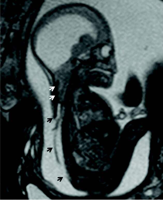
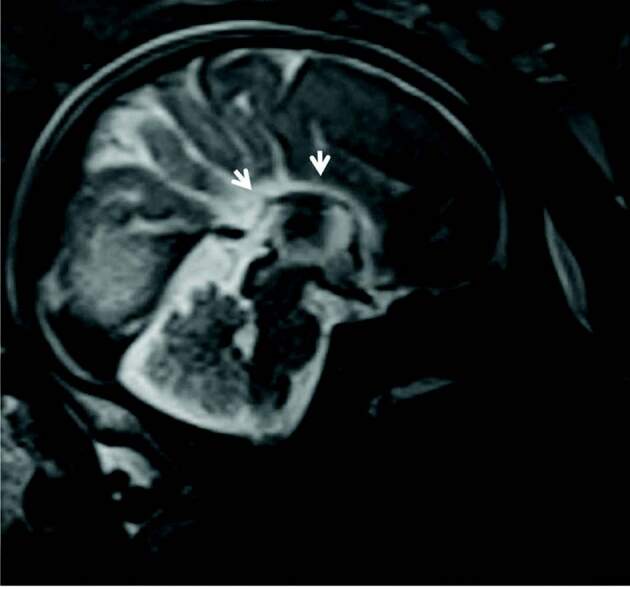
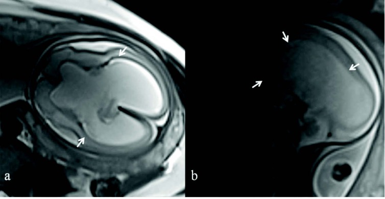
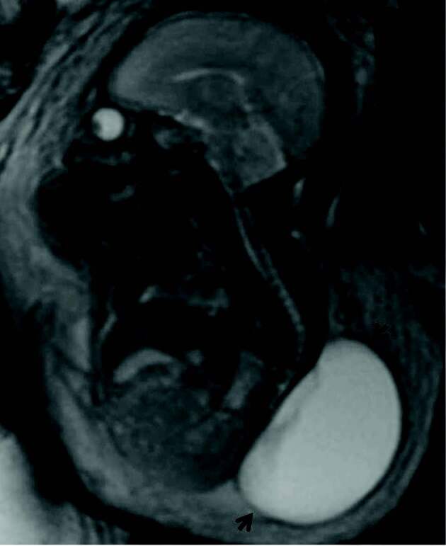
Due to its high resolution and multiplanar imaging capability, fetal MRI contributes significantly to US in the correct prenatal diagnosis of craniospinal anomalies. This contribution especially is significant in neural tube defects, cortical malformations, and ischemic-hemorrhagic lesions.

摘要

背景/目的:通过回顾性比较产前和产后发现,揭示磁共振成像(MRI)在产前诊断胎儿颅脊柱畸形中的超声(US)作用。

材料与方法

在机构审查委员会批准后,我们于 2010 年 1 月至 2020 年 5 月期间,对 301 名妊娠 19-37 周(平均 26.5±6.1 周)的孕妇进行了评估,这些孕妇在胎儿 US 上诊断出颅脊柱异常,随后对其进行 MRI 成像,其中 179 例病例的产前成像结果与产后结果进行了比较。

结果

在 179 名孕妇中,共发现 191 例胎儿颅脊柱畸形。MRI 和 US 的诊断准确率分别为 145(75.9%)和 112(58.6%)。MRI 的诊断性能明显高于 US(p<0.05)。在 53%的病例中,产前 MRI 与产后诊断结果一致。在 28.7%的病例中,产前 MRI 通过改变错误的 US 诊断(8.9%)、显示额外的发现(14%)或确认可疑的 US 诊断(5.8%),从而对 US 做出了贡献。

结论

由于其具有高分辨率和多平面成像能力,胎儿 MRI 对 US 在正确诊断颅脊柱畸形中具有重要作用。这种作用在神经管缺陷、皮质畸形和缺血性-出血性病变中尤为明显。

https://cdn.ncbi.nlm.nih.gov/pmc/blobs/e697/8283491/79e24b2e0697/turkjmedsci-51-1211-fig004.jpg
https://cdn.ncbi.nlm.nih.gov/pmc/blobs/e697/8283491/19049f3512af/turkjmedsci-51-1211-fig001.jpg
https://cdn.ncbi.nlm.nih.gov/pmc/blobs/e697/8283491/d61f7feac559/turkjmedsci-51-1211-fig002.jpg
https://cdn.ncbi.nlm.nih.gov/pmc/blobs/e697/8283491/313932b5e304/turkjmedsci-51-1211-fig003.jpg
https://cdn.ncbi.nlm.nih.gov/pmc/blobs/e697/8283491/79e24b2e0697/turkjmedsci-51-1211-fig004.jpg

相似文献

1
Fetal US and MRI in detection of craniospinal anomalies with postnatal correlation: single-center experience.胎儿 US 和 MRI 在后生性颅脊柱异常检测中的应用:单中心经验。
Turk J Med Sci. 2021 Jun 28;51(3):1211-1219. doi: 10.3906/sag-2011-122.
2
Role of magnetic resonance imaging in fetuses with mild or moderate ventriculomegaly in the era of fetal neurosonography: systematic review and meta-analysis.磁共振成像在胎儿神经超声时代轻度或中度脑室扩张胎儿中的作用:系统评价和荟萃分析。
Ultrasound Obstet Gynecol. 2019 Aug;54(2):164-171. doi: 10.1002/uog.20197. Epub 2019 Jul 11.
3
Fetal Neurosonogaphy: Ultrasound and Magnetic Resonance Imaging in Competition.胎儿神经超声检查:超声与磁共振成像的竞争关系
Ultraschall Med. 2016 Dec;37(6):555-557. doi: 10.1055/s-0042-117142. Epub 2016 Dec 15.
4
Contribution of Magnetic Resonance Imaging to Ultrasound for the Evaluation of Fetal Central Nervous System Anomalies.磁共振成像在超声评估胎儿中枢神经系统异常中的作用
J Reprod Med. 2017 May-Jun;62(5-6):295-9.
5
Role of prenatal magnetic resonance imaging in fetuses with isolated severe ventriculomegaly at neurosonography: A multicenter study.产前磁共振成像在神经超声检查孤立性重度脑室扩张胎儿中的作用:一项多中心研究。
Eur J Obstet Gynecol Reprod Biol. 2021 Dec;267:105-110. doi: 10.1016/j.ejogrb.2021.10.014. Epub 2021 Oct 23.
6
Prenatal diagnosis of congenital head, face, and neck malformations-Is complementary fetal MRI of value?先天性头、面、颈部畸形的产前诊断——胎儿 MRI 检查有价值吗?
Prenat Diagn. 2020 Jan;40(1):142-150. doi: 10.1002/pd.5593. Epub 2019 Nov 11.
7
Detecting Fetal Central Nervous System Anomalies Using Magnetic Resonance Imaging and Ultrasound.磁共振成像与超声联合检测胎儿中枢神经系统异常
Med Arch. 2021 Feb;75(1):45-49. doi: 10.5455/medarh.2021.75.45-49.
8
MRI in the diagnosis of fetal developmental brain abnormalities: the MERIDIAN diagnostic accuracy study.磁共振成像在胎儿发育性脑异常诊断中的应用:MERIDIAN 诊断准确性研究。
Health Technol Assess. 2019 Sep;23(49):1-144. doi: 10.3310/hta23490.
9
Contribution of magnetic resonance imaging to the prenatal diagnosis of common congenital vascular anomalies.磁共振成像在常见先天性血管畸形产前诊断中的应用。
Pediatr Radiol. 2021 Aug;51(9):1626-1636. doi: 10.1007/s00247-021-05031-w. Epub 2021 Apr 23.
10
Prenatal congenital vertical talus (rocker bottom foot): a marker for multisystem anomalies.产前先天性垂直距骨(摇椅底足):多系统异常的一个标志。
Pediatr Radiol. 2017 Dec;47(13):1793-1799. doi: 10.1007/s00247-017-3957-z. Epub 2017 Sep 6.

引用本文的文献

1
Frontonasal Dysplasia: A Diagnostic Challenge with Fetal MRI in Twin Pregnancy.额鼻发育异常:双胎妊娠中胎儿磁共振成像的诊断挑战
Child Neurol Open. 2023 Mar 6;10:2329048X231157147. doi: 10.1177/2329048X231157147. eCollection 2023 Jan-Dec.
2
A single-center experience of magnetic resonance imaging findings of fetal sacrococcygeal teratomas.胎儿骶尾部畸胎瘤的磁共振成像表现:单中心经验
Turk J Med Sci. 2022 Aug;52(4):1190-1196. doi: 10.55730/1300-0144.5423. Epub 2022 Aug 10.
3
Use of magnetic resonance imaging in the diagnosis of fetal vertebral abnormalities in utero: a single-center retrospective cohort study.

本文引用的文献

1
Fetal MRI of the central nervous system: State-of-the-art.中枢神经系统的胎儿磁共振成像:最新技术。
Eur J Radiol. 2017 Aug;93:273-283. doi: 10.1016/j.ejrad.2017.06.004. Epub 2017 Jun 17.
2
Added value of fetal MRI in fetuses with suspected brain abnormalities on neurosonography: a systematic review and meta-analysis.胎儿磁共振成像在超声检查怀疑有脑部异常胎儿中的附加价值:一项系统评价和荟萃分析。
J Matern Fetal Neonatal Med. 2016 Sep;29(18):2949-61. doi: 10.3109/14767058.2015.1109621. Epub 2015 Nov 23.
3
Fetal Brain MRI: Novel Classification and Contribution to Sonography.
磁共振成像在产前诊断胎儿脊柱异常中的应用:一项单中心回顾性队列研究
Quant Imaging Med Surg. 2022 Jun;12(6):3391-3405. doi: 10.21037/qims-21-1070.
胎儿脑部磁共振成像:新的分类及其对超声检查的贡献
Ultraschall Med. 2016 Apr;37(2):176-84. doi: 10.1055/s-0034-1384935. Epub 2014 Dec 23.
4
Is fetal magnetic resonance imaging indicated when ultrasound isolated mild ventriculomegaly is present in pregnancies with no risk factors?在无风险因素的妊娠中,当超声检查发现孤立性轻度脑室扩张时,是否需要进行胎儿磁共振成像检查?
Prenat Diagn. 2012 Aug;32(8):752-7. doi: 10.1002/pd.3896. Epub 2012 May 14.
5
Malformations of cortical development: diagnostic accuracy of fetal MR imaging.皮层发育畸形:胎儿磁共振成像的诊断准确性。
Radiology. 2012 Jun;263(3):843-55. doi: 10.1148/radiol.12102492. Epub 2012 Apr 10.
6
MRI: is there a role in obstetrics?磁共振成像:在产科中有作用吗?
Clin Obstet Gynecol. 2012 Mar;55(1):352-66. doi: 10.1097/GRF.0b013e3182487d04.
7
Neurodevelopment after prenatal diagnosis of isolated agenesis of the corpus callosum: an integrative review.孤立性胼胝体发育不全的产前诊断后的神经发育:综合评价。
Am J Obstet Gynecol. 2012 Apr;206(4):337.e1-5. doi: 10.1016/j.ajog.2011.12.024. Epub 2011 Dec 27.
8
Magnetic resonance imaging of the fetal central nervous system, head, neck, and chest.胎儿中枢神经系统、头部、颈部和胸部的磁共振成像。
Semin Ultrasound CT MR. 2012 Feb;33(1):86-101. doi: 10.1053/j.sult.2011.10.005.
9
Contribution of MRI to ultrasound in the diagnosis of fetal anomalies.MRI 对超声在胎儿畸形诊断中的作用。
J Magn Reson Imaging. 2012 Apr;35(4):882-90. doi: 10.1002/jmri.23502. Epub 2011 Nov 29.
10
Practice guidelines for performance of the routine mid-trimester fetal ultrasound scan.孕中期常规胎儿超声检查操作指南。
Ultrasound Obstet Gynecol. 2011 Jan;37(1):116-26. doi: 10.1002/uog.8831.