Farm Animal Genetic Resources Exploration and Innovation Key Laboratory of Sichuan Province, Chengdu Campus, Sichuan Agricultural University, 611130 Chengdu, Sichuan, China.
Farm Animal Genetic Resources Exploration and Innovation Key Laboratory of Sichuan Province, Chengdu Campus, Sichuan Agricultural University, 611130 Chengdu, Sichuan, China.
Poult Sci. 2021 Feb;100(2):1098-1108. doi: 10.1016/j.psj.2020.10.017. Epub 2020 Oct 21.
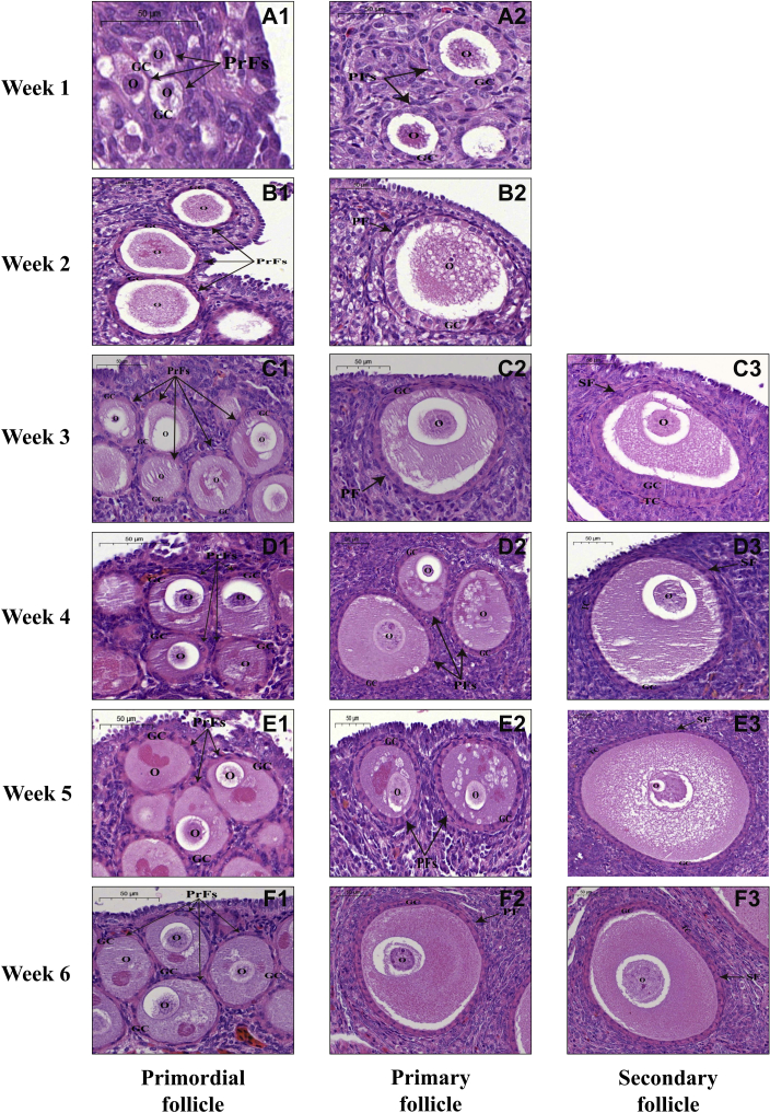
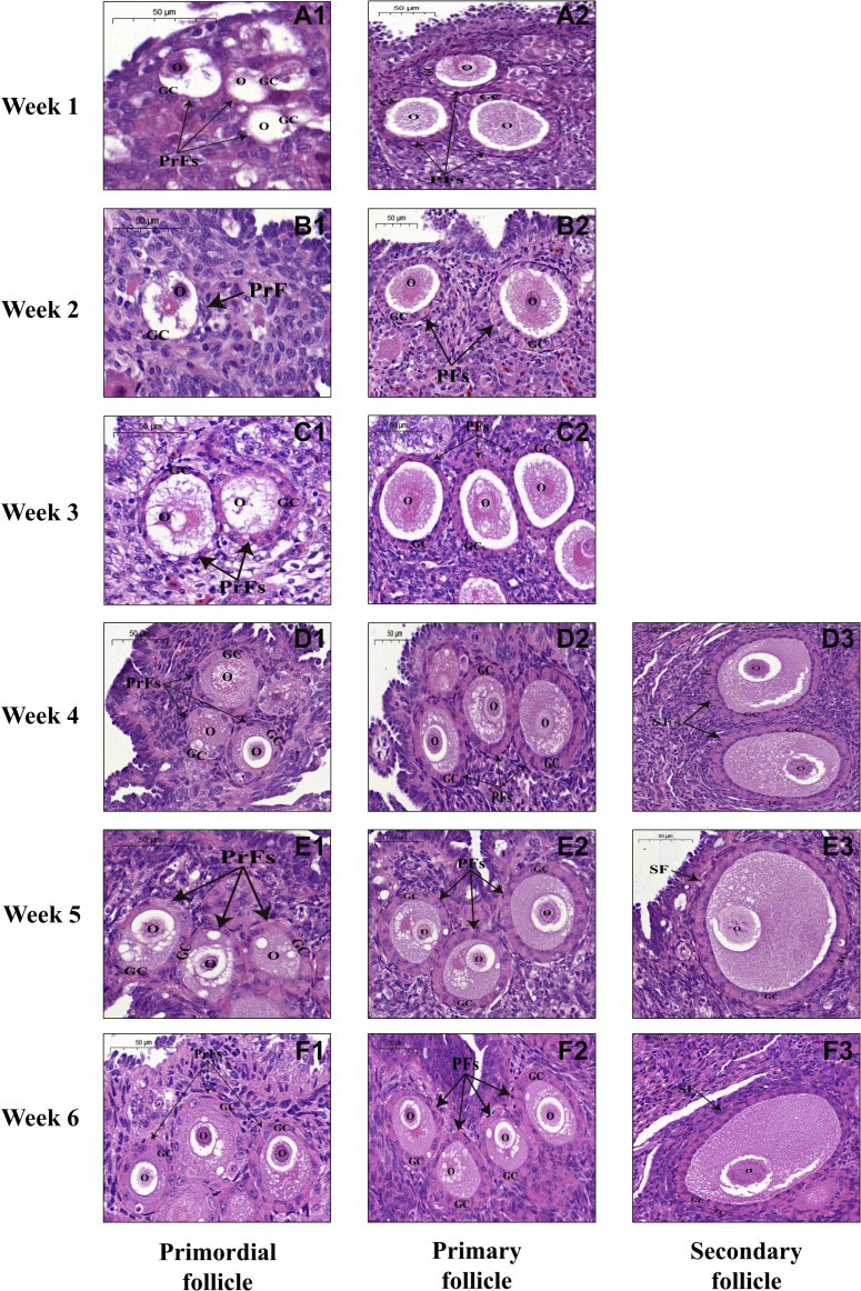
In contrast to the later stages of follicle development, little is known about the characteristics and mechanisms associated with early folliculogenesis in avian species. The objectives of the present study were to examine and compare the histomorphological and molecular changes of primordial, primary, and secondary follicles from duck and goose ovaries during the first 6 post-hatching week. Morphological analysis showed that the length and width of both duck and goose ovaries increased steadily during weeks 1 to 5 but increased acutely at week 6, whereas a greater increment was observed in the ovarian length of ducks than that of geese during weeks 4 to 5. Furthermore, smaller diameters of the 3 categories of follicles were observed in ducks than those in geese at the first appearance, but they reached a similar size at week 6. More importantly, secondary follicles were found in the ovaries of ducks 1 wk earlier than in those of geese. These results indicated a more rapid growth rate for ovarian follicles in ducks than in geese during early post-hatching development. At the molecular level, it was found that the mRNAs encoding follicle stimulating hormone receptor (FSHR), anti-Müllerian hormone (AMH), B-cell leukemia/lymphoma 2, and cysteine-dependent aspartate specific protease 3 (CASPASE3) were ubiquitously expressed in all ovarian follicles of ducks and geese with different expression profiles in each follicular category during the first 6 post-hatching week. Notably, transcript levels of FSHR, AMH, and CASPASE3 changed differently between ducks and geese during weeks 5 to 6, which was postulated to be one of the mechanisms inducing more rapid growth of ovarian follicles in ducks rather than in geese. In conclusion, our results revealed, for the first time, differences in early folliculogenesis, including the rate of growth of each follicular category and the timing of transition of primary to secondary follicles, between ducks and geese, and these differences could result from different expression profiles of FSHR, AMH, and CASPASE3 during early post-hatching development.
与卵泡发育后期相比,人们对禽类早期卵泡发生的特征和机制知之甚少。本研究旨在研究和比较鸭和鹅卵巢在孵化后第 1 至 6 周内原始卵泡、初级卵泡和次级卵泡的组织形态学和分子变化。形态学分析表明,鸭和鹅的卵巢长度和宽度在第 1 周到第 5 周期间稳步增加,但在第 6 周急剧增加,而在第 4 周到第 5 周期间,鸭的卵巢长度增加幅度大于鹅。此外,在首次出现时,鸭的这 3 类卵泡的直径小于鹅,但在第 6 周时达到相似大小。更重要的是,鸭的次级卵泡比鹅的更早出现在卵巢中。这些结果表明,在孵化后早期发育过程中,鸭的卵巢卵泡生长速度快于鹅。在分子水平上,发现鸭和鹅的所有卵巢卵泡中均广泛表达卵泡刺激素受体(FSHR)、抗苗勒管激素(AMH)、B 细胞白血病/淋巴瘤 2 和半胱氨酸依赖性天冬氨酸特异性蛋白酶 3(CASPASE3)的 mRNA,并且在孵化后第 1 周内每种卵泡类别中都有不同的表达谱。值得注意的是,FSHR、AMH 和 CASPASE3 的转录水平在第 5 周到第 6 周期间在鸭和鹅之间发生了不同的变化,这被认为是导致鸭的卵巢卵泡生长速度快于鹅的机制之一。总之,我们的研究结果首次揭示了鸭和鹅之间早期卵泡发生的差异,包括每个卵泡类别的生长速度和初级卵泡向次级卵泡的转变时间,这些差异可能是由于 FSHR、AMH 和 CASPASE3 在孵化后早期发育过程中的不同表达谱所致。