Thibodeau Ryan, Jafroodifar Abtin, Quraeshi Sahir, Lisi Michele
Department of Radiology, SUNY Upstate Medical University, 750 East Adams Street, Syracuse, NY 13210, USA.
Radiol Case Rep. 2021 Apr;16(4):753-759. doi: 10.1016/j.radcr.2021.01.025. Epub 2021 Jan 15.
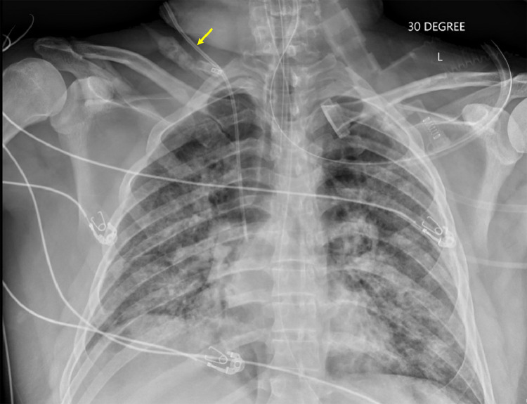
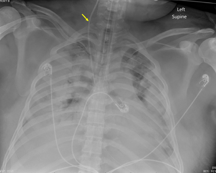
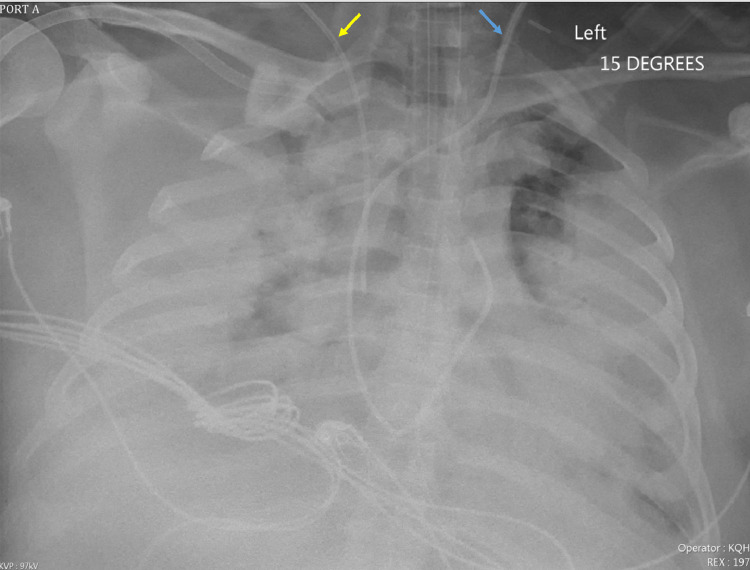
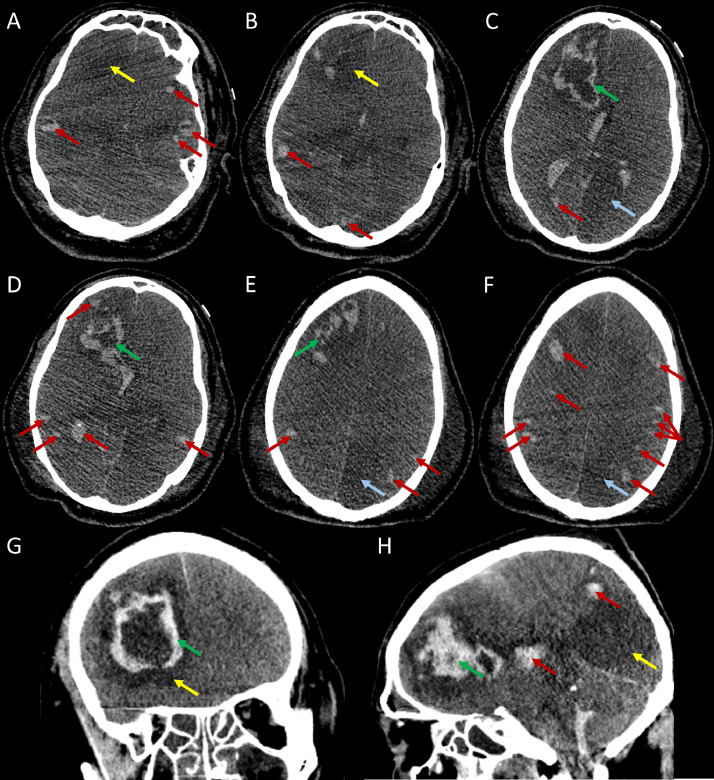
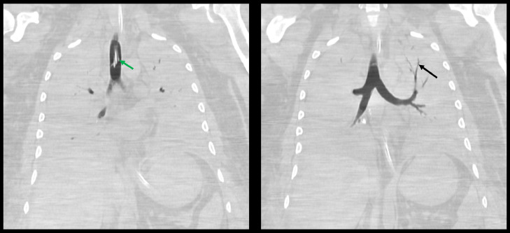
Coronavirus disease 2019 (COVID-19) may present with pulmonary and extrapulmonary manifestations. We present a 41-year-old patient who presented with 1 week of increasing dyspnea and fever and initial chest radiography demonstrated bilateral diffuse infiltrates. Due to the patient's progressively worsening symptoms, he was intubated, paralyzed, sedated. He began proning, 100% fractional inspired oxygenation ventilation, and veno-venous extracorporeal membrane oxygenation. Computed tomography of the thorax revealed completely opacified lungs bilaterally with the exception of a small, aerated apicoposterior segment of the left lung. Computed tomography of the head demonstrated several areas of hemorrhage, areas of hypodensity consistent with posterior cerebral artery and middle cerebral artery territory infarcts, and findings consistent with transtentorial herniation. Given the radiologic findings and nonprogressive clinical status, the family placed the patient on comfort care and the patient died within minutes of extubation. As with our patient at the time of admission, presenting symptoms and clinical laboratory data provide reliable prognostic factors for patients with COVID-19.
2019冠状病毒病(COVID-19)可能有肺部和肺外表现。我们报告一名41岁患者,出现进行性加重的呼吸困难和发热1周,初始胸部X线检查显示双侧弥漫性浸润。由于患者症状逐渐恶化,他被插管、麻痹、镇静。他开始俯卧位通气、100%纯氧通气,并进行静脉-静脉体外膜肺氧合。胸部计算机断层扫描显示双侧肺完全实变,左肺尖后段有一小片充气区域除外。头部计算机断层扫描显示多个出血区域、与大脑后动脉和大脑中动脉供血区梗死相符的低密度区域,以及与小脑幕切迹疝相符的表现。鉴于影像学检查结果和病情无进展,家属让患者接受舒适护理,患者在拔管后几分钟内死亡。与我们的患者入院时情况一样,出现的症状和临床实验室数据为COVID-19患者提供了可靠的预后因素。