Athinoula A. Martinos Center for Biomedical Imaging, Department of Radiology, Massachusetts General Hospital, 149 Thirteenth Street, Suite, 2301, Charlestown 02129, MA, United States; Department of Radiology, Harvard Medical School, Boston, MA, United States.
Athinoula A. Martinos Center for Biomedical Imaging, Department of Radiology, Massachusetts General Hospital, 149 Thirteenth Street, Suite, 2301, Charlestown 02129, MA, United States; Department of Neurology, Ruijin Hospital & Shanghai Jiao Tong University School of Medicine, Shanghai, China.
Neuroimage. 2021 Apr 15;230:117807. doi: 10.1016/j.neuroimage.2021.117807. Epub 2021 Jan 29.
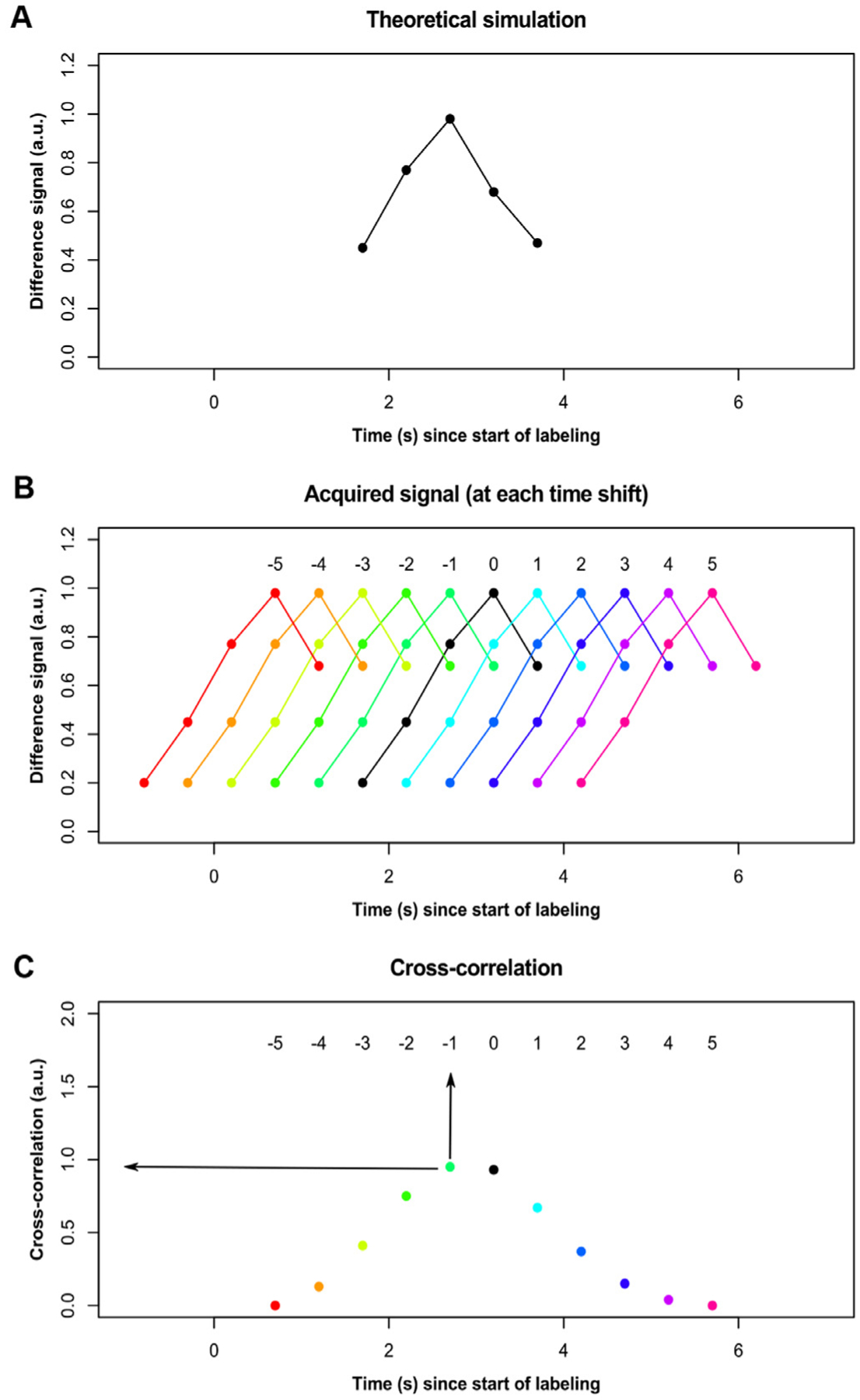
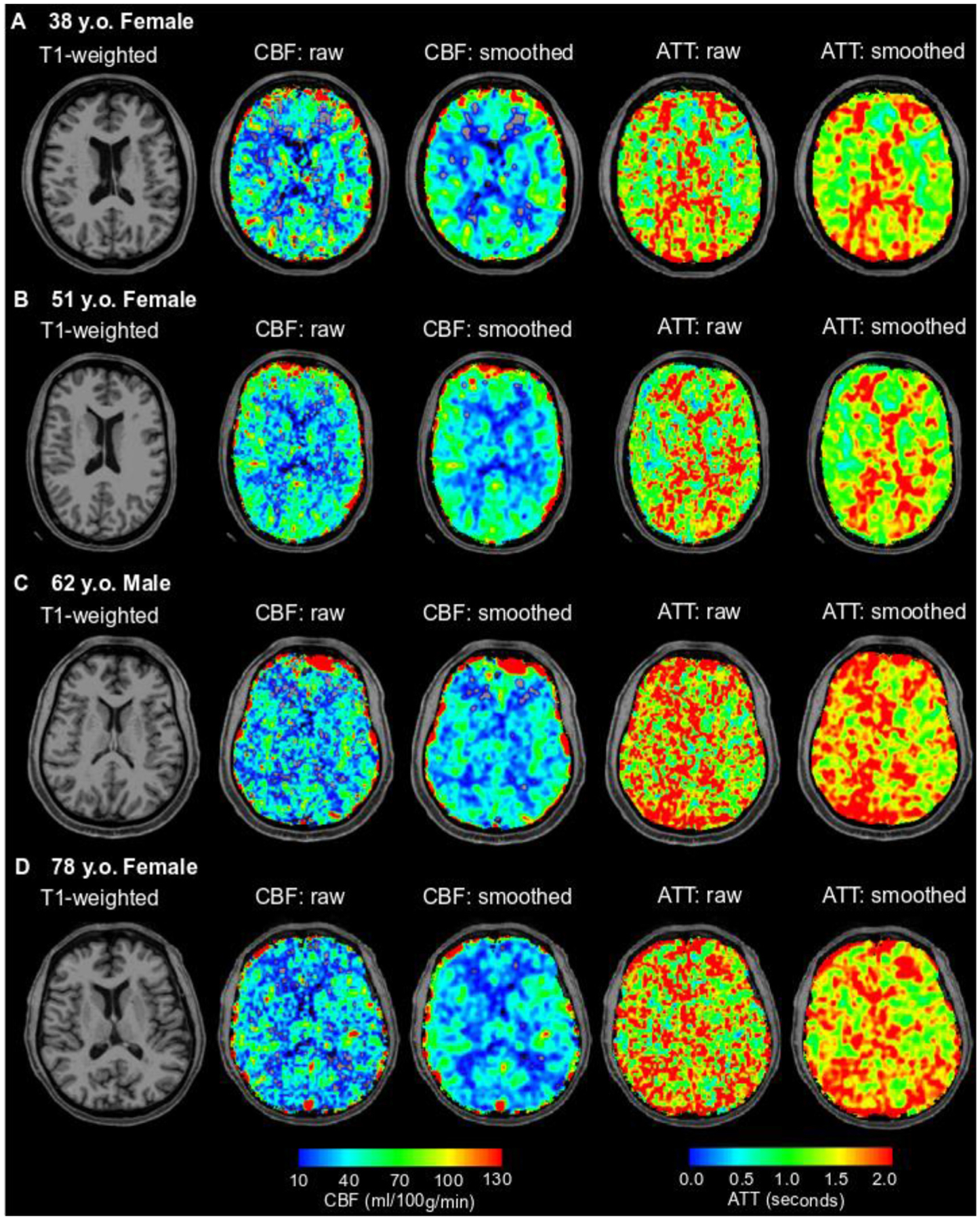
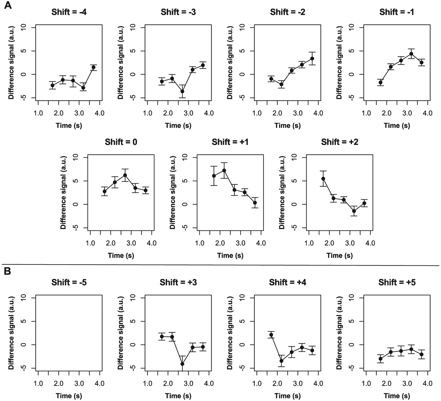
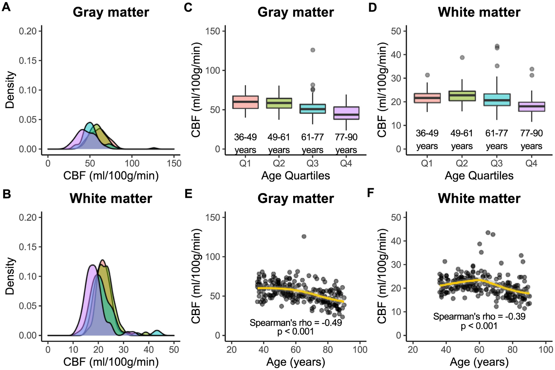
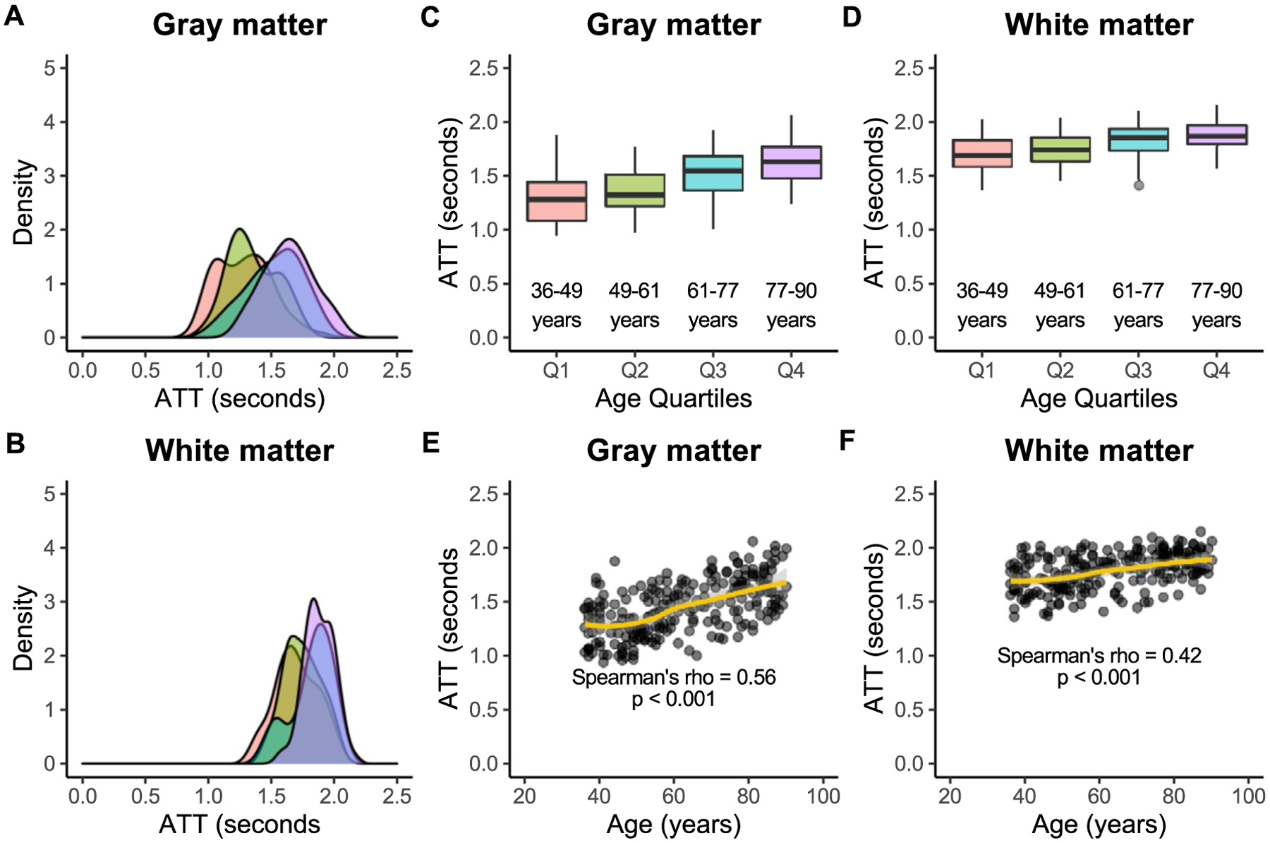
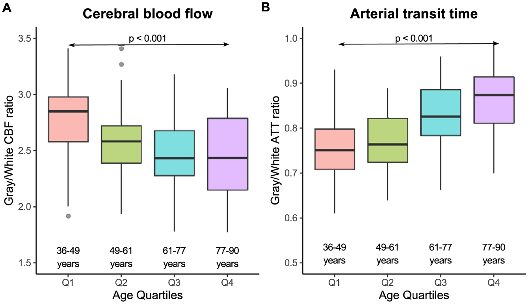
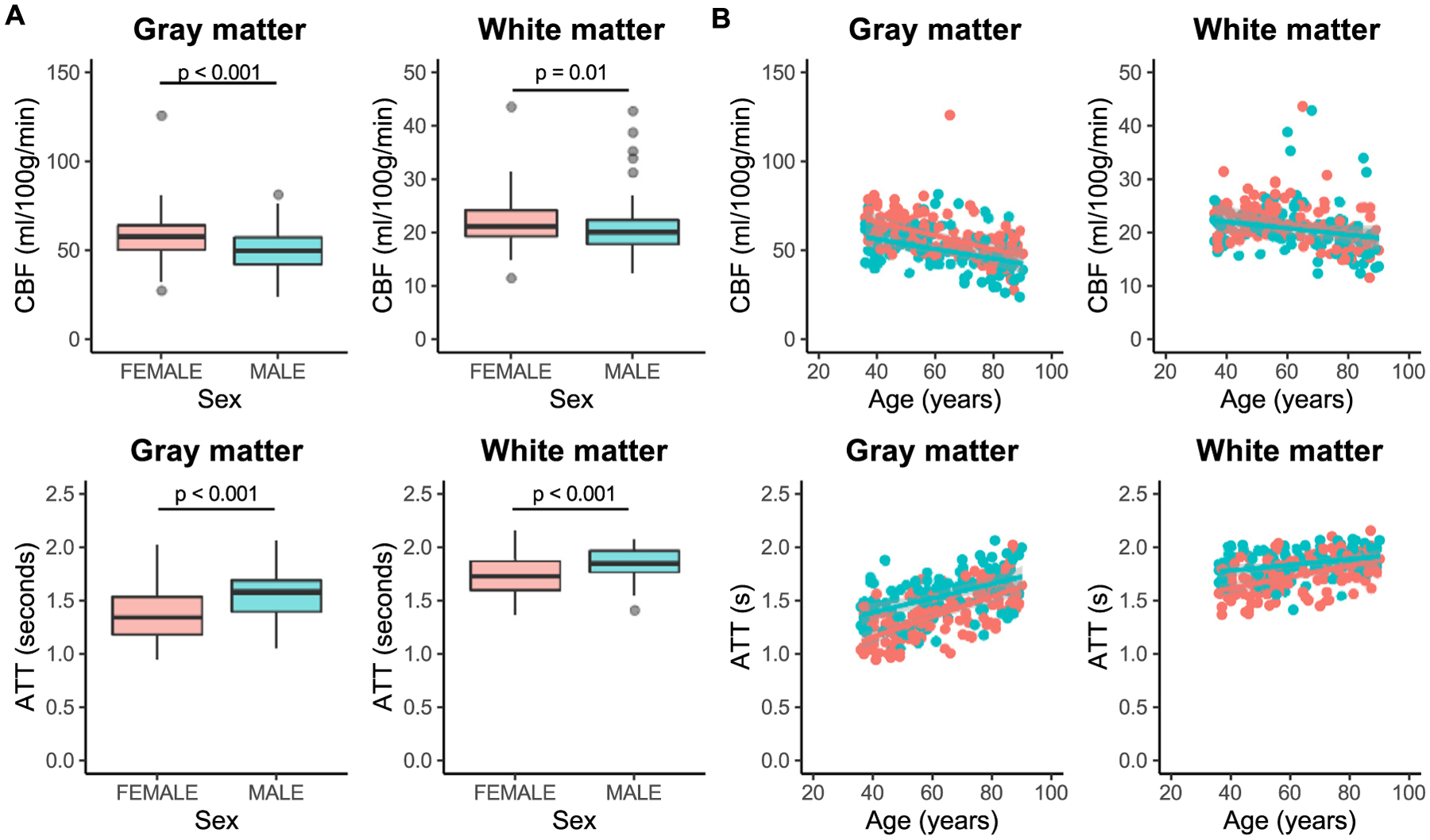
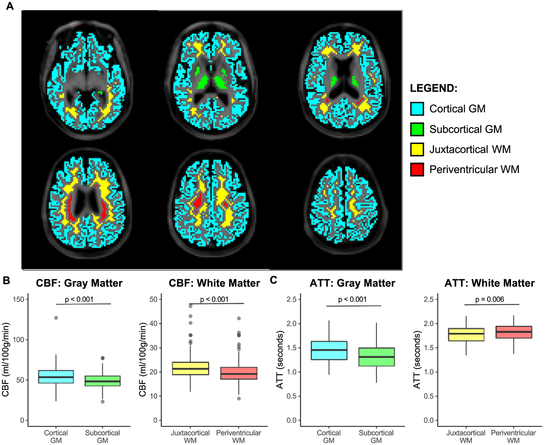
Arterial spin labeling (ASL) magnetic resonance imaging (MRI) has become a popular approach for studying cerebral hemodynamics in a range of disorders and has recently been included as part of the Human Connectome Project-Aging (HCP-A). Due to the high spatial resolution and multiple post-labeling delays, ASL data from HCP-A holds promise for localization of hemodynamic signals not only in gray matter but also in white matter. However, gleaning information about white matter hemodynamics with ASL is challenging due in part to longer blood arrival times in white matter compared to gray matter. In this work, we present an analytical approach for deriving measures of cerebral blood flow (CBF) and arterial transit times (ATT) from the ASL data from HCP-A and report on gray and white matter hemodynamics in a large cohort (n = 234) of typically aging adults (age 36-90 years). Pseudo-continuous ASL data were acquired with labeling duration = 1500 ms and five post-labeling delays = 200 ms, 700 ms, 1200, 1700 ms, and 2200 ms. ATT values were first calculated on a voxel-wise basis through normalized cross-correlation analysis of the acquired signal time course in that voxel and an expected time course based on an acquisition-specific Bloch simulation. CBF values were calculated using a two-compartment model and with age-appropriate blood water longitudinal relaxation times. Using this approach, we found that white matter CBF reduces (ρ = 0.39) and white matter ATT elongates (ρ = 0.42) with increasing age (p < 0.001). In addition, CBF is lower and ATTs are longer in white matter compared to gray matter across the adult lifespan (Wilcoxon signed-rank tests; p < 0.001). We also found sex differences with females exhibiting shorter white matter ATTs than males, independently of age (Wilcoxon rank-sum test; p < 0.001). Finally, we have shown that CBF and ATT values are spatially heterogeneous, with significant differences in cortical versus subcortical gray matter and juxtacortical versus periventricular white matter. These results serve as a characterization of normative physiology across the human lifespan against which hemodynamic impairment due to cerebrovascular or neurodegenerative diseases could be compared in future studies.
动脉自旋标记 (ASL) 磁共振成像 (MRI) 已成为研究一系列疾病中脑血流动力学的一种流行方法,最近已被纳入人类连接组计划-衰老 (HCP-A) 的一部分。由于具有高空间分辨率和多个后标记延迟,因此 HCP-A 的 ASL 数据有望不仅在灰质中,而且在白质中定位血流信号。然而,由于与灰质相比,白质中的血液到达时间更长,因此用 ASL 来获取关于白质血流动力学的信息具有挑战性。在这项工作中,我们提出了一种从 HCP-A 的 ASL 数据中推导出脑血流量 (CBF) 和动脉传输时间 (ATT) 的分析方法,并报告了一大群(n=234)典型衰老成年人(年龄 36-90 岁)的灰质和白质血流动力学。采用伪连续 ASL 数据,标记持续时间 = 1500ms,5 个后标记延迟 = 200ms、700ms、1200ms、1700ms 和 2200ms。通过在该体素中获取的信号时间过程与基于特定采集的 Bloch 模拟的期望时间过程的归一化互相关分析,首先在体素基础上计算 ATT 值。使用双室模型并使用与年龄相关的血液水纵向弛豫时间来计算 CBF 值。使用这种方法,我们发现白质 CBF 随着年龄的增加而降低(ρ=0.39),白质 ATT 随着年龄的增加而延长(ρ=0.42)(p<0.001)。此外,在整个成年期,白质的 CBF 较低,ATT 较长,与灰质相比(Wilcoxon 符号秩检验;p<0.001)。我们还发现了性别差异,女性的白质 ATT 比男性短,与年龄无关(Wilcoxon 秩和检验;p<0.001)。最后,我们表明 CBF 和 ATT 值具有空间异质性,皮质与皮质下灰质以及皮质旁与脑室周围白质之间存在显著差异。这些结果是对人类整个生命周期正常生理学的描述,未来的研究可以将其与脑血管或神经退行性疾病引起的血流动力学障碍进行比较。