• 文献检索
  • 文档翻译
  • 深度研究
  • 学术资讯
  • Suppr Zotero 插件Zotero 插件
  • 邀请有礼
  • 套餐&价格
  • 历史记录
应用&插件
Suppr Zotero 插件Zotero 插件浏览器插件Mac 客户端Windows 客户端微信小程序
定价
高级版会员购买积分包购买API积分包
服务
文献检索文档翻译深度研究API 文档MCP 服务
关于我们
关于 Suppr公司介绍联系我们用户协议隐私条款
关注我们

Suppr 超能文献

核心技术专利:CN118964589B侵权必究
粤ICP备2023148730 号-1Suppr @ 2026

文献检索

告别复杂PubMed语法,用中文像聊天一样搜索,搜遍4000万医学文献。AI智能推荐,让科研检索更轻松。

立即免费搜索

文件翻译

保留排版,准确专业,支持PDF/Word/PPT等文件格式,支持 12+语言互译。

免费翻译文档

深度研究

AI帮你快速写综述,25分钟生成高质量综述,智能提取关键信息,辅助科研写作。

立即免费体验

肌球蛋白结合蛋白 C 的 N 端在完整的心肌中朝向肌动蛋白丝延伸。

The N terminus of myosin-binding protein C extends toward actin filaments in intact cardiac muscle.

机构信息

Department of Molecular Physiology and Biophysics, Cardiovascular Research Institute, University of Vermont, Burlington, VT.

Division of Cell Biology and Imaging, Department of Radiology, University of Massachusetts Medical School, Worcester, MA.

出版信息

J Gen Physiol. 2021 Mar 1;153(3). doi: 10.1085/jgp.202012726.

DOI:10.1085/jgp.202012726
PMID:33528507
原文链接:https://pmc.ncbi.nlm.nih.gov/articles/PMC7852460/
Abstract

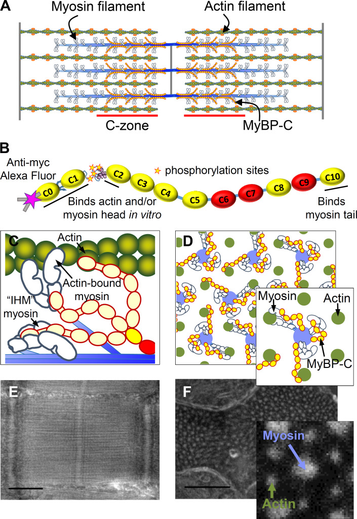
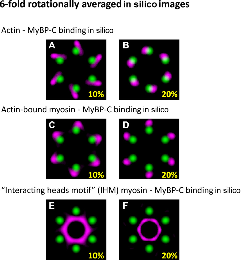
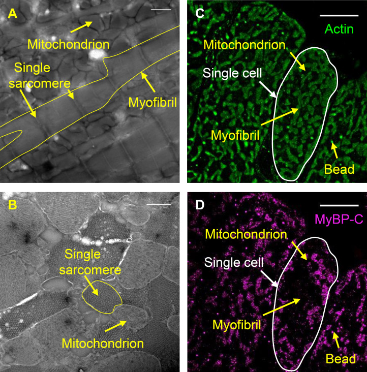
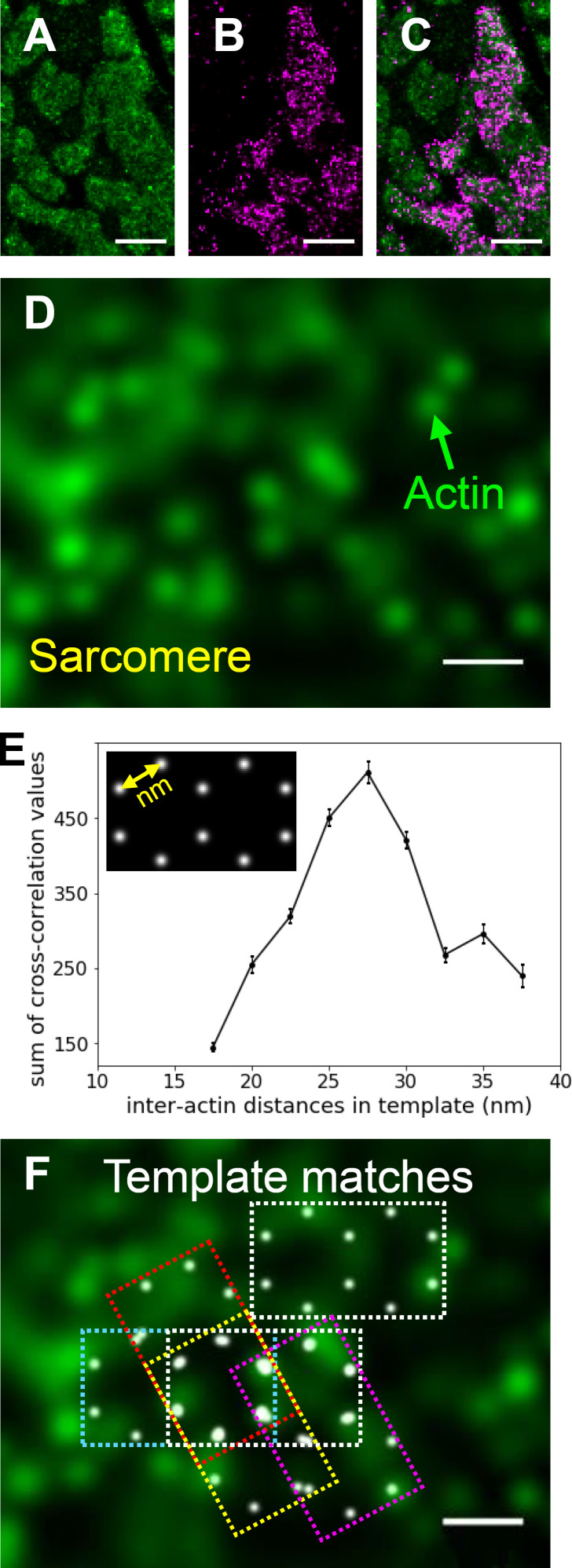
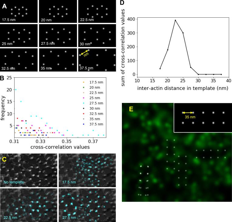
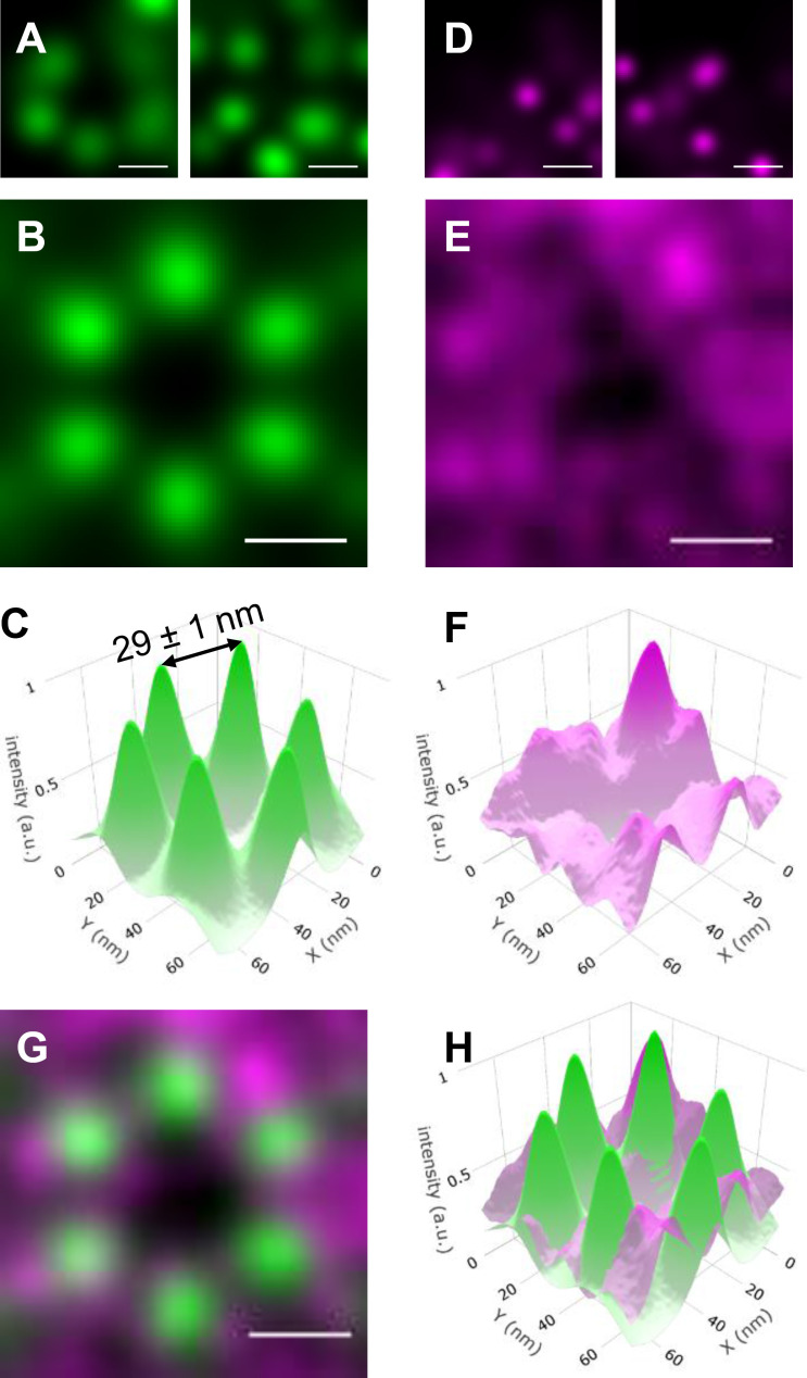
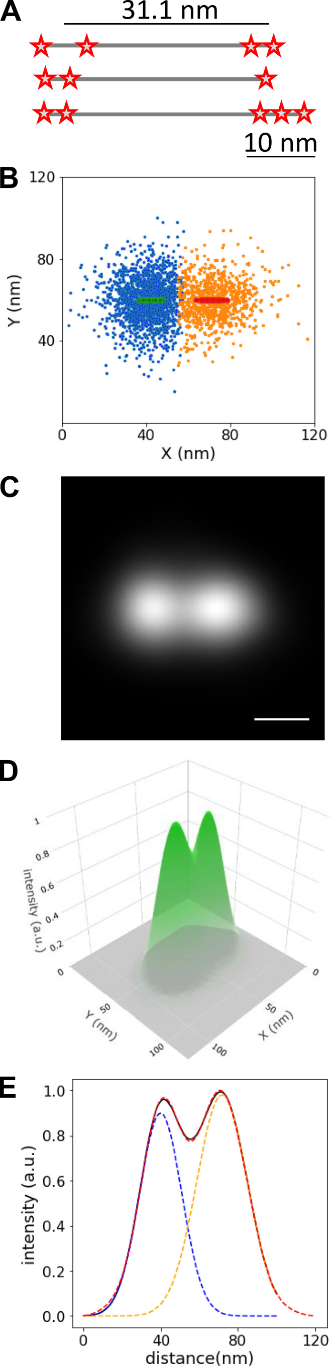
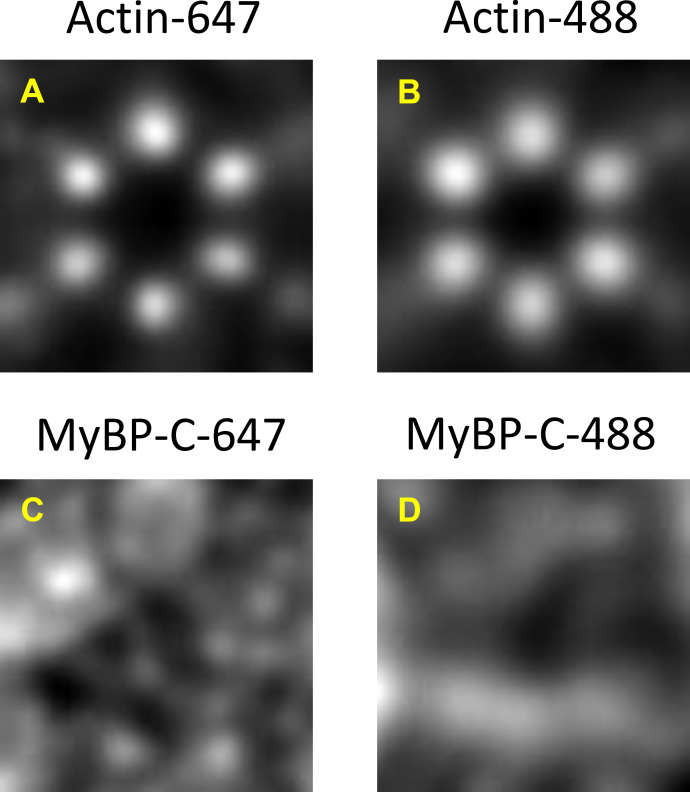
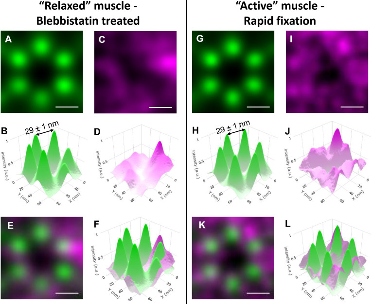
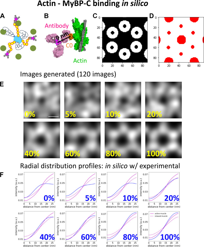
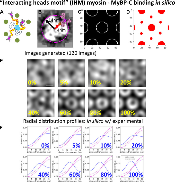
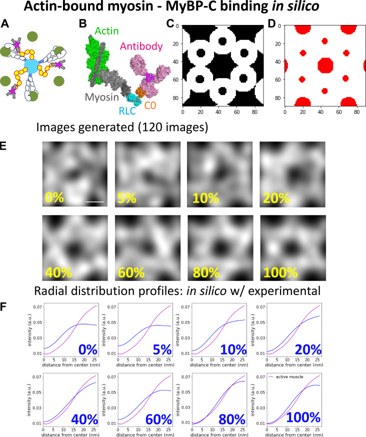
Myosin and actin filaments are highly organized within muscle sarcomeres. Myosin-binding protein C (MyBP-C) is a flexible, rod-like protein located within the C-zone of the sarcomere. The C-terminal domain of MyBP-C is tethered to the myosin filament backbone, and the N-terminal domains are postulated to interact with actin and/or the myosin head to modulate filament sliding. To define where the N-terminal domains of MyBP-C are localized in the sarcomere of active and relaxed mouse myocardium, the relative positions of the N terminus of MyBP-C and actin were imaged in fixed muscle samples using super-resolution fluorescence microscopy. The resolution of the imaging was enhanced by particle averaging. The images demonstrate that the position of the N terminus of MyBP-C is biased toward the actin filaments in both active and relaxed muscle preparations. Comparison of the experimental images with images generated in silico, accounting for known binding partner interactions, suggests that the N-terminal domains of MyBP-C may bind to actin and possibly the myosin head but only when the myosin head is in the proximity of an actin filament. These physiologically relevant images help define the molecular mechanism by which the N-terminal domains of MyBP-C may search for, and capture, molecular binding partners to tune cardiac contractility.

摘要

肌球蛋白和肌动蛋白丝在肌肉肌节内高度组织化。肌球蛋白结合蛋白 C(MyBP-C)是一种位于肌节 C 带的灵活的杆状蛋白。MyBP-C 的 C 端结构域与肌球蛋白丝主干相连,而 N 端结构域则被假设与肌动蛋白和/或肌球蛋白头部相互作用,以调节丝滑动。为了确定 MyBP-C 的 N 端结构域在活跃和松弛的小鼠心肌肌节中的定位,使用超分辨率荧光显微镜在固定的肌肉样本中对 MyBP-C 的 N 端和肌动蛋白的相对位置进行成像。通过粒子平均提高了成像的分辨率。这些图像表明,在活跃和松弛的肌肉标本中,MyBP-C 的 N 端位置偏向肌动蛋白丝。将实验图像与考虑已知结合伴侣相互作用的计算图像进行比较表明,MyBP-C 的 N 端结构域可能与肌动蛋白结合,并且可能与肌球蛋白头部结合,但只有当肌球蛋白头部靠近肌动蛋白丝时才会结合。这些与生理相关的图像有助于定义 MyBP-C 的 N 端结构域可能寻找和捕获分子结合伴侣以调节心脏收缩性的分子机制。

https://cdn.ncbi.nlm.nih.gov/pmc/blobs/122c/7852460/e9a470a27fb6/JGP_202012726_FigS10.jpg
https://cdn.ncbi.nlm.nih.gov/pmc/blobs/122c/7852460/5819d2567f9e/JGP_202012726_Fig1.jpg
https://cdn.ncbi.nlm.nih.gov/pmc/blobs/122c/7852460/b4bc5c756ee0/JGP_202012726_FigS1.jpg
https://cdn.ncbi.nlm.nih.gov/pmc/blobs/122c/7852460/61740beb210f/JGP_202012726_FigS2.jpg
https://cdn.ncbi.nlm.nih.gov/pmc/blobs/122c/7852460/cda15649941d/JGP_202012726_Fig2.jpg
https://cdn.ncbi.nlm.nih.gov/pmc/blobs/122c/7852460/0c8640b6173b/JGP_202012726_FigS3.jpg
https://cdn.ncbi.nlm.nih.gov/pmc/blobs/122c/7852460/17291e5a4916/JGP_202012726_Fig3.jpg
https://cdn.ncbi.nlm.nih.gov/pmc/blobs/122c/7852460/2e481542e647/JGP_202012726_FigS4.jpg
https://cdn.ncbi.nlm.nih.gov/pmc/blobs/122c/7852460/43b1c0b5a32c/JGP_202012726_FigS5.jpg
https://cdn.ncbi.nlm.nih.gov/pmc/blobs/122c/7852460/d06dec2eadf0/JGP_202012726_FigS6.jpg
https://cdn.ncbi.nlm.nih.gov/pmc/blobs/122c/7852460/ca9a11bcbfb7/JGP_202012726_Fig4.jpg
https://cdn.ncbi.nlm.nih.gov/pmc/blobs/122c/7852460/46fffcac03ca/JGP_202012726_FigS7.jpg
https://cdn.ncbi.nlm.nih.gov/pmc/blobs/122c/7852460/12b567eba2c1/JGP_202012726_FigS8.jpg
https://cdn.ncbi.nlm.nih.gov/pmc/blobs/122c/7852460/5b22ffdd4f78/JGP_202012726_FigS9.jpg
https://cdn.ncbi.nlm.nih.gov/pmc/blobs/122c/7852460/e9a470a27fb6/JGP_202012726_FigS10.jpg

相似文献

1
The N terminus of myosin-binding protein C extends toward actin filaments in intact cardiac muscle.肌球蛋白结合蛋白 C 的 N 端在完整的心肌中朝向肌动蛋白丝延伸。
J Gen Physiol. 2021 Mar 1;153(3). doi: 10.1085/jgp.202012726.
2
Cryo-electron tomography of intact cardiac muscle reveals myosin binding protein-C linking myosin and actin filaments.冷冻电镜断层扫描完整的心肌肌球蛋白结合蛋白-C 连接肌球蛋白和肌动蛋白丝。
J Muscle Res Cell Motil. 2023 Sep;44(3):165-178. doi: 10.1007/s10974-023-09647-3. Epub 2023 Apr 28.
3
Structural evidence for the interaction of C-protein (MyBP-C) with actin and sequence identification of a possible actin-binding domain.C蛋白(肌球蛋白结合蛋白C,MyBP-C)与肌动蛋白相互作用的结构证据及一个可能的肌动蛋白结合结构域的序列鉴定。
J Mol Biol. 2003 Aug 15;331(3):713-24. doi: 10.1016/s0022-2836(03)00781-2.
4
Earning stripes: myosin binding protein-C interactions with actin.获得条纹:肌球蛋白结合蛋白-C 与肌动蛋白的相互作用。
Pflugers Arch. 2014 Mar;466(3):445-50. doi: 10.1007/s00424-013-1432-8. Epub 2014 Jan 19.
5
Functional role of myosin-binding protein H in thick filaments of developing vertebrate fast-twitch skeletal muscle.肌球蛋白结合蛋白 H 在脊椎动物快肌发育中的粗肌丝中的功能作用。
J Gen Physiol. 2024 Dec 2;156(12). doi: 10.1085/jgp.202413604. Epub 2024 Oct 7.
6
Structure, interactions and function of the N-terminus of cardiac myosin binding protein C (MyBP-C): who does what, with what, and to whom?心肌肌球蛋白结合蛋白 C(MyBP-C)N 端的结构、相互作用和功能:谁与谁、与什么以及与谁相互作用?
J Muscle Res Cell Motil. 2012 May;33(1):83-94. doi: 10.1007/s10974-012-9291-z. Epub 2012 Apr 20.
7
Skeletal MyBP-C isoforms tune the molecular contractility of divergent skeletal muscle systems.骨骼肌球蛋白结合蛋白-C 的同工型调节不同骨骼肌肉系统的分子收缩性。
Proc Natl Acad Sci U S A. 2019 Oct 22;116(43):21882-21892. doi: 10.1073/pnas.1910549116. Epub 2019 Oct 7.
8
MYBPC3 truncation mutations enhance actomyosin contractile mechanics in human hypertrophic cardiomyopathy.MYBPC3 截断突变增强人类肥厚型心肌病中的肌球蛋白收缩力学。
J Mol Cell Cardiol. 2019 Feb;127:165-173. doi: 10.1016/j.yjmcc.2018.12.003. Epub 2018 Dec 11.
9
Electron microscopy and 3D reconstruction of F-actin decorated with cardiac myosin-binding protein C (cMyBP-C).用心脏肌球蛋白结合蛋白 C(cMyBP-C)装饰的 F-肌动蛋白的电子显微镜和 3D 重建。
J Mol Biol. 2011 Jul 8;410(2):214-25. doi: 10.1016/j.jmb.2011.05.010. Epub 2011 May 13.
10
The C-terminal IgI domains of myosin-binding proteins C and H (MyBP-C and MyBP-H) are both necessary and sufficient for the intracellular crosslinking of sarcomeric myosin in transfected non-muscle cells.肌球蛋白结合蛋白C和H(MyBP-C和MyBP-H)的C端免疫球蛋白样结构域对于转染的非肌肉细胞中肌节肌球蛋白的细胞内交联而言既是必需的也是充分的。
J Cell Sci. 2002 Sep 1;115(Pt 17):3517-26. doi: 10.1242/jcs.115.17.3517.

引用本文的文献

1
Hypertrophic cardiomyopathy: insights into pathophysiology and novel therapeutic strategies from clinical studies.肥厚型心肌病:临床研究对病理生理学及新型治疗策略的见解
Egypt Heart J. 2025 Jan 7;77(1):5. doi: 10.1186/s43044-024-00600-4.
2
Mechanism-based myofilament manipulation to treat diastolic dysfunction in HFpEF.基于机制的肌丝调控以治疗射血分数保留的心力衰竭中的舒张功能障碍。
Front Physiol. 2024 Dec 12;15:1512550. doi: 10.3389/fphys.2024.1512550. eCollection 2024.
3
Functional role of myosin-binding protein H in thick filaments of developing vertebrate fast-twitch skeletal muscle.

本文引用的文献

1
Imaging ATP Consumption in Resting Skeletal Muscle: One Molecule at a Time.静息骨骼肌中ATP消耗的成像:逐个分子研究
Biophys J. 2020 Sep 15;119(6):1050-1055. doi: 10.1016/j.bpj.2020.07.036. Epub 2020 Aug 15.
2
The hypertrophic cardiomyopathy mutations R403Q and R663H increase the number of myosin heads available to interact with actin.肥厚型心肌病突变 R403Q 和 R663H 增加了与肌动蛋白相互作用的肌球蛋白头部数量。
Sci Adv. 2020 Apr 3;6(14):eaax0069. doi: 10.1126/sciadv.aax0069. eCollection 2020 Apr.
3
Myosin filament-based regulation of the dynamics of contraction in heart muscle.
肌球蛋白结合蛋白 H 在脊椎动物快肌发育中的粗肌丝中的功能作用。
J Gen Physiol. 2024 Dec 2;156(12). doi: 10.1085/jgp.202413604. Epub 2024 Oct 7.
4
Reassessing the unifying hypothesis for hypercontractility caused by myosin mutations in hypertrophic cardiomyopathy.重新评估肥厚型心肌病肌球蛋白突变引起的高收缩性的统一假说。
EMBO J. 2024 Oct;43(19):4139-4155. doi: 10.1038/s44318-024-00199-x. Epub 2024 Aug 27.
5
Functional role of myosin-binding protein H in thick filaments of developing vertebrate fast-twitch skeletal muscle.肌球蛋白结合蛋白H在发育中的脊椎动物快肌骨骼肌粗肌丝中的功能作用。
bioRxiv. 2024 May 13:2024.05.10.593199. doi: 10.1101/2024.05.10.593199.
6
Bringing into focus the central domains C3-C6 of myosin binding protein C.聚焦肌球蛋白结合蛋白C的中心结构域C3-C6。
Front Physiol. 2024 Feb 29;15:1370539. doi: 10.3389/fphys.2024.1370539. eCollection 2024.
7
Cardiac myosin-binding protein C N-terminal interactions with myosin and actin filaments: Opposite effects of phosphorylation and M-domain mutations.心肌球蛋白结合蛋白 C N 端与肌球蛋白和肌动蛋白丝的相互作用:磷酸化和 M 结构域突变的相反影响。
J Mol Cell Cardiol. 2024 Jan;186:125-137. doi: 10.1016/j.yjmcc.2023.11.010. Epub 2023 Nov 24.
8
Visualization of cardiac thick filament dynamics in ex vivo heart preparations.离体心脏标本中心脏粗丝动力学的可视化。
J Mol Cell Cardiol. 2023 Dec;185:88-98. doi: 10.1016/j.yjmcc.2023.10.013. Epub 2023 Nov 2.
9
Cryo-electron tomography of intact cardiac muscle reveals myosin binding protein-C linking myosin and actin filaments.冷冻电镜断层扫描完整的心肌肌球蛋白结合蛋白-C 连接肌球蛋白和肌动蛋白丝。
J Muscle Res Cell Motil. 2023 Sep;44(3):165-178. doi: 10.1007/s10974-023-09647-3. Epub 2023 Apr 28.
10
The contribution of N-terminal truncated cMyBPC to in vivo cardiac function.N 端截短型 cMyBPC 对体内心脏功能的贡献。
J Gen Physiol. 2023 Jun 5;155(6). doi: 10.1085/jgp.202213318. Epub 2023 Apr 17.
基于肌球蛋白丝的心肌收缩动力学调节。
Proc Natl Acad Sci U S A. 2020 Apr 7;117(14):8177-8186. doi: 10.1073/pnas.1920632117. Epub 2020 Mar 27.
4
Site-specific phosphorylation of myosin binding protein-C coordinates thin and thick filament activation in cardiac muscle.肌球蛋白结合蛋白-C 的位点特异性磷酸化协调心肌中细肌丝和粗肌丝的激活。
Proc Natl Acad Sci U S A. 2019 Jul 30;116(31):15485-15494. doi: 10.1073/pnas.1903033116. Epub 2019 Jul 15.
5
Cardiac myosin binding protein-C phosphorylation regulates the super-relaxed state of myosin.肌球蛋白结合蛋白-C 磷酸化调节肌球蛋白的超松弛状态。
Proc Natl Acad Sci U S A. 2019 Jun 11;116(24):11731-11736. doi: 10.1073/pnas.1821660116. Epub 2019 May 29.
6
MYBPC3 truncation mutations enhance actomyosin contractile mechanics in human hypertrophic cardiomyopathy.MYBPC3 截断突变增强人类肥厚型心肌病中的肌球蛋白收缩力学。
J Mol Cell Cardiol. 2019 Feb;127:165-173. doi: 10.1016/j.yjmcc.2018.12.003. Epub 2018 Dec 11.
7
N-Terminal Domains of Cardiac Myosin Binding Protein C Cooperatively Activate the Thin Filament.心肌球蛋白结合蛋白 C 的 N 端结构域协同激活细肌丝。
Structure. 2018 Dec 4;26(12):1604-1611.e4. doi: 10.1016/j.str.2018.08.007. Epub 2018 Sep 27.
8
Hypertrophic cardiomyopathy and the myosin mesa: viewing an old disease in a new light.肥厚型心肌病与肌球蛋白台座:以新视角看待一种古老疾病。
Biophys Rev. 2018 Feb;10(1):27-48. doi: 10.1007/s12551-017-0274-6. Epub 2017 Jul 17.
9
The myosin mesa and the basis of hypercontractility caused by hypertrophic cardiomyopathy mutations.肌球蛋白台面与肥厚型心肌病突变导致的高收缩性基础。
Nat Struct Mol Biol. 2017 Jun;24(6):525-533. doi: 10.1038/nsmb.3408. Epub 2017 May 8.
10
Structure of actomyosin rigour complex at 5.2 Å resolution and insights into the ATPase cycle mechanism.肌球蛋白刚性复合物的结构分辨率为 5.2Å 及对 ATP 酶循环机制的深入了解。
Nat Commun. 2017 Jan 9;8:13969. doi: 10.1038/ncomms13969.