Nuzzi Raffaele, Buono Lola, Scalabrin Simona, De Iuliis Marco, Bussolati Benedetta
Eye Clinic, Department of Surgical Sciences, University of Turin, AOU Città della Salute e della Scienza, Turin, Italy.
Department of Biotechnology and Health Sciences, University of Turin, Turin, Italy.
Stem Cells Int. 2021 Jan 16;2021:6644463. doi: 10.1155/2021/6644463. eCollection 2021.
Human corneal endothelial cells (HCECs) are essential to visual function; however, since they have limited proliferative capacity , they are prone to corneal endothelial dysfunction. At present, the only treatment is a corneal transplantation from donor cadavers. Also, due to a global shortage of donor corneas, it is important to find alternative strategies. Recent studies highlight that stem cell-derived extracellular vesicles (EVs) play a relevant role in stem cell-induced regeneration by reprogramming injured cells and inducing proregenerative pathways. The aim of this work is to evaluate whether EVs derived from mesenchymal stem cells (MSC-EVs) are able to promote regeneration of damaged HCECs.
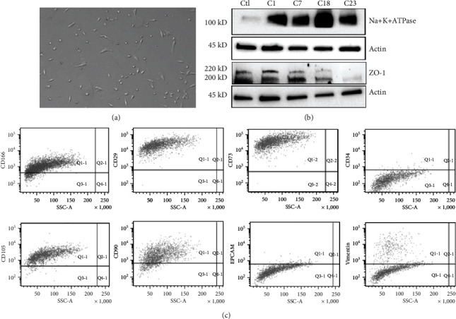
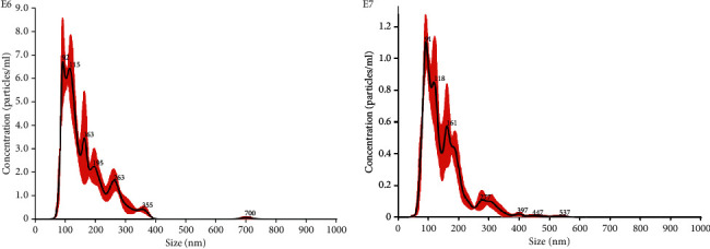
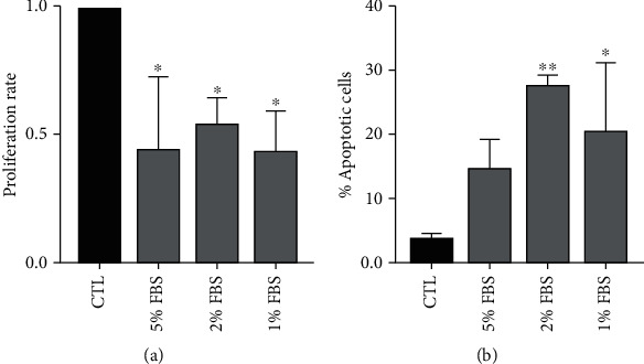
We isolated HCECs from discarded corneas in patients undergoing corneal transplantation or enucleation ( = 23 patients). Bone marrow mesenchymal stem cells (MSCs) were obtained from Lonza, cultured, and characterized. MSC-EVs were obtained from supernatants of MSCs. In order to establish a valid damage model to test the regenerative potential of EVs on HCECs, we evaluated the proliferation rate and the apoptosis after exposing the cells to serum-deprived medium at different concentrations for 24 hours. We then evaluated the HCEC migration through a wound healing assay.
In the selected serum deprivation damage conditions, the treatment with different doses of MSC-EVs resulted in a significantly higher proliferation rate of HCECs at all the tested concentrations of EVs (5-20 × 10 MSC-EV/cell). MSC-EVs/cell induced a significant decrease in number of total apoptotic cells after 24 hours of serum deprivation. Finally, the wound healing assay showed a significantly faster repair of the wound after HCEC treatment with MSC-EVs.
Results highlight the already well-known proregenerative potential of MSC-EVs in a totally new biological model, the endothelium of the cornea. MSC-EVs, indeed, induced proliferation and survival of HCECs, promoting the migration of HCECs .
人角膜内皮细胞(HCECs)对视觉功能至关重要;然而,由于它们的增殖能力有限,容易发生角膜内皮功能障碍。目前,唯一的治疗方法是使用供体尸体的角膜移植。此外,由于全球供体角膜短缺,寻找替代策略很重要。最近的研究表明,干细胞衍生的细胞外囊泡(EVs)通过重编程受损细胞和诱导促再生途径,在干细胞诱导的再生中发挥相关作用。这项工作的目的是评估间充质干细胞衍生的细胞外囊泡(MSC-EVs)是否能够促进受损HCECs的再生。
我们从接受角膜移植或眼球摘除术的患者(n = 23例)废弃的角膜中分离出HCECs。从Lonza公司获得骨髓间充质干细胞(MSCs),进行培养和鉴定。从MSCs的上清液中获得MSC-EVs。为了建立一个有效的损伤模型来测试EVs对HCECs的再生潜力,我们评估了细胞在不同浓度的血清饥饿培养基中暴露24小时后的增殖率和凋亡情况。然后,我们通过伤口愈合试验评估HCEC的迁移。
在选定的血清饥饿损伤条件下,用不同剂量的MSC-EVs处理后,在所有测试的EVs浓度(5 - 20×10个MSC-EV/细胞)下,HCECs的增殖率均显著更高。在血清饥饿24小时后,MSC-EVs/细胞诱导总凋亡细胞数量显著减少。最后,伤口愈合试验表明,用MSC-EVs处理HCECs后,伤口修复明显加快。
结果突出了MSC-EVs在一个全新的生物学模型——角膜内皮中早已为人所知的促再生潜力。事实上,MSC-EVs诱导了HCECs的增殖和存活,促进了HCECs的迁移。