Chen Junyu, Sivan Unnikrishnan, Tan Sin Lih, Lippo Luciana, De Angelis Jessica, Labella Rossella, Singh Amit, Chatzis Alexandros, Cheuk Stanley, Medhghalchi Mino, Gil Jesus, Hollander Georg, Marsden Brian D, Williams Richard, Ramasamy Saravana K, Kusumbe Anjali P
Tissue and Tumor Microenvironments Group, Kennedy Institute of Rheumatology, University of Oxford, Oxford OX3 7FY, UK.
Department of Prosthodontics, State Key Laboratory of Oral Diseases, West China Hospital of Stomatology, Sichuan University, Chengdu 610041, China.
Sci Adv. 2021 Feb 3;7(6). doi: 10.1126/sciadv.abd7819. Print 2021 Feb.
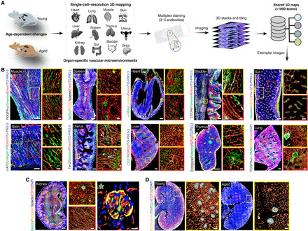
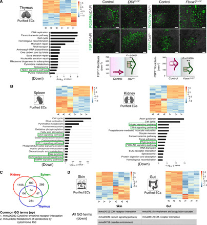
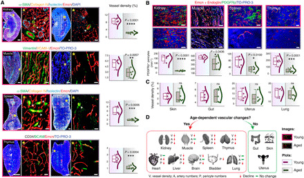
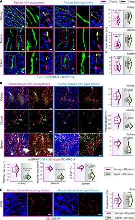
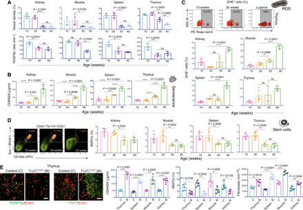
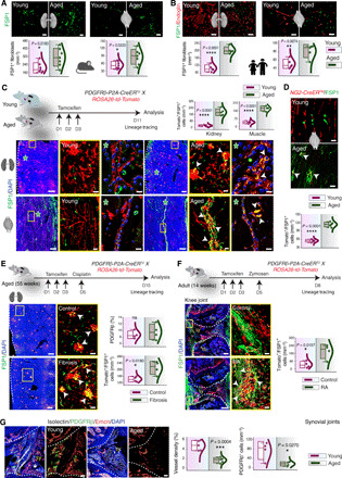
Blood vessels provide supportive microenvironments for maintaining tissue functions. Age-associated vascular changes and their relation to tissue aging and pathology are poorly understood. Here, we perform 3D imaging of young and aging vascular beds. Multiple organs in mice and humans demonstrate an age-dependent decline in vessel density and pericyte numbers, while highly remodeling tissues such as skin preserve the vasculature. Vascular attrition precedes the appearance of cellular hallmarks of aging such as senescence. Endothelial VEGFR2 loss-of-function mice demonstrate that vascular perturbations are sufficient to stimulate cellular changes coupled with aging. Age-associated tissue-specific molecular changes in the endothelium drive vascular loss and dictate pericyte to fibroblast differentiation. Lineage tracing of perivascular cells with inducible PDGFRβ and NG2 Cre mouse lines demonstrated that increased pericyte to fibroblast differentiation distinguishes injury-induced organ fibrosis and zymosan-induced arthritis. To spur further discoveries, we provide a freely available resource with 3D vascular and tissue maps.
血管为维持组织功能提供支持性微环境。与年龄相关的血管变化及其与组织衰老和病理学的关系尚不清楚。在此,我们对年轻和衰老的血管床进行三维成像。小鼠和人类的多个器官显示血管密度和周细胞数量随年龄下降,而皮肤等高度重塑的组织则保留了脉管系统。血管损耗先于衰老的细胞特征如衰老的出现。内皮细胞VEGFR2功能丧失的小鼠表明血管扰动足以刺激与衰老相关的细胞变化。内皮细胞中与年龄相关的组织特异性分子变化驱动血管损失,并决定周细胞向成纤维细胞的分化。使用诱导型PDGFRβ和NG2 Cre小鼠品系对血管周围细胞进行谱系追踪表明,周细胞向成纤维细胞分化的增加区分了损伤诱导的器官纤维化和酵母聚糖诱导的关节炎。为推动进一步的发现,我们提供了一个免费的资源,其中包含三维血管和组织图谱。