Department of Neuroscience, Central Clinical School, Monash University, Prahran, VIC, 3004, Australia.
Faculty of Applied Medical Sciences, Taif University, Taif, 26521, Kingdom of Saudi Arabia.
Sci Rep. 2021 Feb 3;11(1):2890. doi: 10.1038/s41598-021-82346-6.
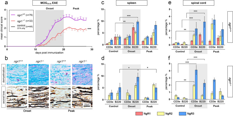
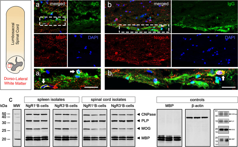
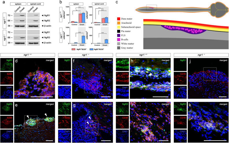
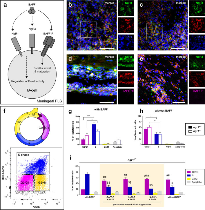
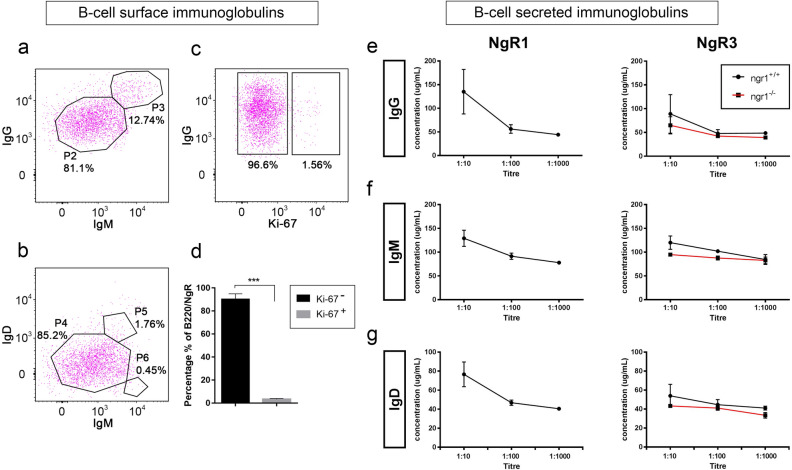
We have previously reported evidence that Nogo-A activation of Nogo-receptor 1 (NgR1) can drive axonal dystrophy during the neurological progression of experimental autoimmune encephalomyelitis (EAE). However, the B-cell activating factor (BAFF/BlyS) may also be an important ligand of NgR during neuroinflammation. In the current study we define that NgR1 and its homologs may contribute to immune cell signaling during EAE. Meningeal B-cells expressing NgR1 and NgR3 were identified within the lumbosacral spinal cords of ngr1 EAE-induced mice at clinical score 1. Furthermore, increased secretion of immunoglobulins that bound to central nervous system myelin were shown to be generated from isolated NgR1- and NgR3-expressing B-cells of ngr1 EAE-induced mice. In vitro BAFF stimulation of NgR1- and NgR3-expressing B cells, directed them into the cell cycle DNA synthesis phase. However, when we antagonized BAFF signaling by co-incubation with recombinant BAFF-R, NgR1-Fc, or NgR3 peptides, the B cells remained in the G0/G1 phase. The data suggest that B cells express NgR1 and NgR3 during EAE, being localized to infiltrates of the meninges and that their regulation is governed by BAFF signaling.
我们之前的研究表明,Nogo-A 对 Nogo 受体 1(NgR1)的激活可在实验性自身免疫性脑脊髓炎(EAE)的神经进展过程中导致轴突萎缩。然而,B 细胞激活因子(BAFF/BlyS)在神经炎症期间也可能是 NgR 的重要配体。在本研究中,我们定义 NgR1 及其同源物可能在 EAE 期间参与免疫细胞信号传导。在临床评分 1 时,在 ngr1 EAE 诱导的小鼠的腰骶脊髓中鉴定出表达 NgR1 和 NgR3 的脑膜 B 细胞。此外,从 ngr1 EAE 诱导的小鼠中分离出表达 NgR1 和 NgR3 的 B 细胞,显示出与中枢神经系统髓鞘结合的免疫球蛋白的增加分泌。体外 BAFF 刺激表达 NgR1 和 NgR3 的 B 细胞,将其导向细胞周期 DNA 合成期。然而,当我们通过与重组 BAFF-R、NgR1-Fc 或 NgR3 肽共孵育来拮抗 BAFF 信号时,B 细胞仍处于 G0/G1 期。数据表明,B 细胞在 EAE 期间表达 NgR1 和 NgR3,定位于脑膜浸润处,其调节受 BAFF 信号的控制。