Theotokis Paschalis, Touloumi Olga, Lagoudaki Roza, Nousiopoulou Evangelia, Kesidou Evangelia, Siafis Spyridon, Tselios Theodoros, Lourbopoulos Athanasios, Karacostas Dimitrios, Grigoriadis Nikolaos, Simeonidou Constantina
B' Department of Neurology, Laboratory of Experimental Neurology and Neuroimmunology, AHEPA University Hospital, Aristotle University of Thessaloniki, Stilponos Kiriakides str. 1, 546 36, Thessaloniki, Central Macedonia, Greece.
Department of Chemistry, University of Patras, Rion, 265 04, Patras, Greece.
J Neuroinflammation. 2016 Oct 11;13(1):265. doi: 10.1186/s12974-016-0730-4.
Nogo-A and its putative receptor NgR are considered to be among the inhibitors of axonal regeneration in the CNS. However, few studies so far have addressed the issue of local NgR complex multilateral localization within inflammation in an MS mouse model of autoimmune demyelination.
Chronic experimental autoimmune encephalomyelitis (EAE) was induced in C57BL/6 mice. Analyses were performed on acute (days 18-22) and chronic (day 50) time points and compared to controls. The temporal and spatial expression of the Nogo receptor complex (NgR and coreceptors) was studied at the spinal cord using epifluorescent and confocal microscopy or real-time PCR. Data are expressed as cells/mm, as mean % ± SEM, or as arbitrary units of integrated density.
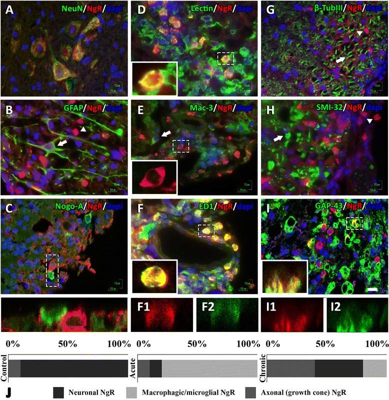
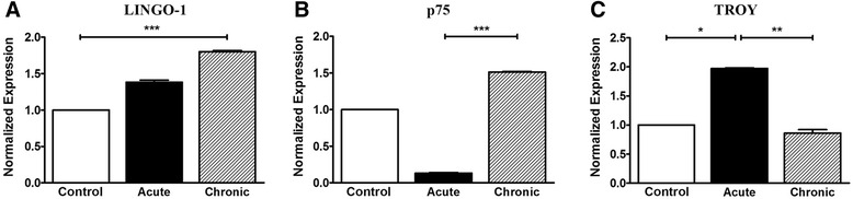
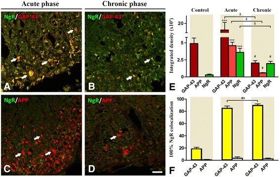
Animals developed a moderate to severe EAE without mortality, followed by a progressive, chronic clinical course. NgR complex spatial expression varied during the main time points of EAE. NgR with coreceptors LINGO-1 and TROY was increased in the spinal cord in the acute phase whereas LINGO-1 and p75 signal seemed to be dominant in the chronic phase, respectively. NgR was detected on gray matter NeuN neurons of the spinal cord, within the white matter inflammatory foci (14.2 ± 4.3 % NgR inflammatory cells), and found to be colocalized with GAP-43 axonal growth cones while no β-TubIII, SMI-32, or APP axons were found as NgR. Among the NgR inflammatory cells, 75.6 ± 9.0 % were microglial/macrophages (lectin), 49.6 ± 14.2 % expressed CD68 (phagocytic ED1 cells), and no cells were Mac-3. Of these macrophages/monocytes, only Arginase-1/NgR but not iNOS/NgR were present in lesions both in acute and chronic phases.
Our data describe in detail the expression of the Nogo receptor complex within the autoimmune inflammatory foci and suggest a possible immune action for NgR apart from the established inhibitory one on axonal growth. Its expression by inflammatory macrophages/monocytes could signify a possible role of these cells on axonal guidance and clearance of the lesioned area during inflammatory demyelination.
Nogo-A及其假定受体NgR被认为是中枢神经系统轴突再生的抑制剂。然而,迄今为止,在自身免疫性脱髓鞘的多发性硬化症小鼠模型中,很少有研究探讨炎症中局部NgR复合物多边定位的问题。
在C57BL/6小鼠中诱导慢性实验性自身免疫性脑脊髓炎(EAE)。在急性(第18 - 22天)和慢性(第50天)时间点进行分析,并与对照组进行比较。使用落射荧光显微镜和共聚焦显微镜或实时PCR研究脊髓中Nogo受体复合物(NgR和共受体)的时空表达。数据以细胞/mm、平均百分比±标准误或积分密度的任意单位表示。
动物出现中度至重度EAE且无死亡,随后是进行性慢性临床病程。NgR复合物的空间表达在EAE的主要时间点有所变化。急性期脊髓中NgR与共受体LINGO-1和TROY增加,而慢性期LINGO-1和p75信号似乎分别占主导。在脊髓灰质NeuN神经元上、白质炎症灶内检测到NgR(14.2±4.3%的NgR炎症细胞),并发现其与GAP-43轴突生长锥共定位,而未发现β-TubIII、SMI-32或APP轴突为NgR。在NgR炎症细胞中,75.6±9.0%为小胶质细胞/巨噬细胞(凝集素),49.6±14.2%表达CD68(吞噬性ED1细胞),且无细胞为Mac-3。在这些巨噬细胞/单核细胞中,急性期和慢性期病变中均仅存在精氨酸酶-1/NgR,而不存在诱导型一氧化氮合酶/NgR。
我们的数据详细描述了Nogo受体复合物在自身免疫性炎症灶中的表达,并提示NgR除了对轴突生长具有既定的抑制作用外,可能还具有免疫作用。炎症巨噬细胞/单核细胞对其表达可能意味着这些细胞在炎症性脱髓鞘过程中对轴突导向和损伤区域清除可能发挥作用。