Department of Nuclear Medicine, University of Würzburg, Würzburg, Germany.
Nuclear Medicine, Medical Faculty, University of Augsburg, Augsburg, Germany.
Eur J Nucl Med Mol Imaging. 2021 Aug;48(9):2761-2770. doi: 10.1007/s00259-020-05170-6. Epub 2021 Feb 4.
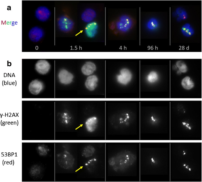
One therapy option for prostate cancer patients with bone metastases is the use of [Ra]RaCl. The α-emitter Ra creates DNA damage tracks along α-particle trajectories (α-tracks) in exposed cells that can be revealed by immunofluorescent staining of γ-H2AX+53BP1 DNA double-strand break markers. We investigated the time- and absorbed dose-dependency of the number of α-tracks in peripheral blood mononuclear cells (PBMCs) of patients undergoing their first therapy with [Ra]RaCl.
Multiple blood samples from nine prostate cancer patients were collected before and after administration of [Ra]RaCl, up to 4 weeks after treatment. γ-H2AX- and 53BP1-positive α-tracks were microscopically quantified in isolated and immuno-stained PBMCs.
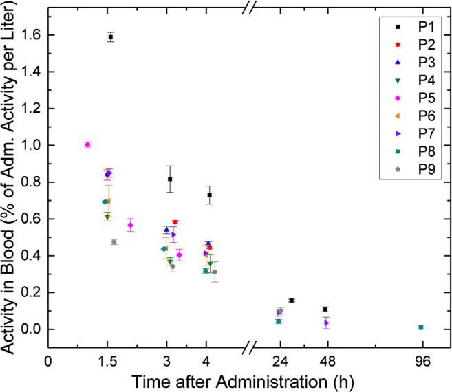
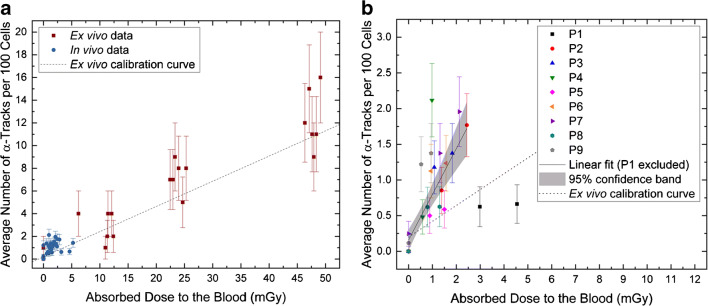
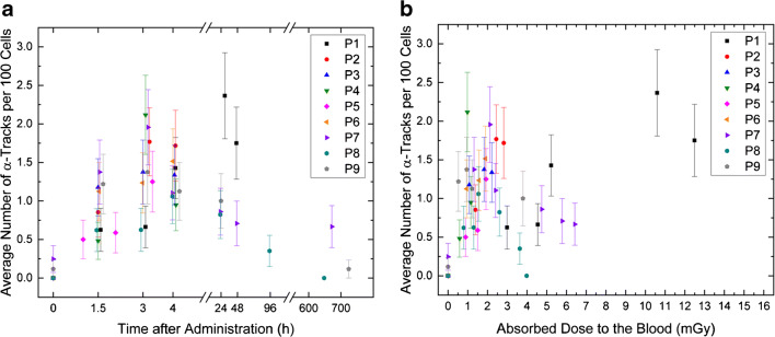
The absorbed doses to the blood were less than 6 mGy up to 4 h after administration and maximally 16 mGy in total. Up to 4 h after administration, the α-track frequency was significantly increased relative to baseline and correlated with the absorbed dose to the blood in the dose range < 3 mGy. In most of the late samples (24 h - 4 weeks after administration), the α-track frequency remained elevated.
The γ-H2AX+53BP1 assay is a potent method for detection of α-particle-induced DNA damages during treatment with or after accidental incorporation of radionuclides even at low absorbed doses. It may serve as a biomarker discriminating α- from β-emitters based on damage geometry.
对于患有骨转移的前列腺癌患者,一种治疗选择是使用 [Ra]RaCl。α 放射体 Ra 在暴露细胞中沿着 α 粒子轨迹(α 轨迹)产生 DNA 损伤轨迹,可以通过 γ-H2AX+53BP1 DNA 双链断裂标记的免疫荧光染色来揭示。我们研究了首次接受 [Ra]RaCl 治疗的患者外周血单核细胞(PBMC)中 α 轨迹数量随时间和吸收剂量的变化。
在给予 [Ra]RaCl 前后,从 9 名前列腺癌患者中采集了多个血液样本,治疗后最多 4 周。在分离和免疫染色的 PBMC 中,通过显微镜定量 γ-H2AX-和 53BP1-阳性的 α 轨迹。
给药后 4 小时内血液吸收剂量小于 6mGy,总剂量最大为 16mGy。给药后 4 小时内,α 轨迹频率相对于基线显著增加,并且与血液吸收剂量呈正相关,吸收剂量范围为<3mGy。在大多数晚期样本(给药后 24 小时至 4 周)中,α 轨迹频率仍然升高。
γ-H2AX+53BP1 测定是一种在治疗期间或放射性核素意外摄入后检测 α 粒子诱导的 DNA 损伤的有效方法,即使在低吸收剂量下也是如此。它可以作为基于损伤几何形状区分 α 辐射体和 β 辐射体的生物标志物。