Kalagiri Rajasree, Stanfield Robyn L, Meisenhelder Jill, La Clair James J, Fuhs Stephen R, Wilson Ian A, Hunter Tony
Molecular and Cell Biology Laboratory, Salk Institute for Biological Studies, La Jolla, CA 92037.
Department of Integrative Structural and Computational Biology, The Scripps Research Institute, La Jolla, CA 92037.
Proc Natl Acad Sci U S A. 2021 Feb 9;118(6). doi: 10.1073/pnas.2010644118.
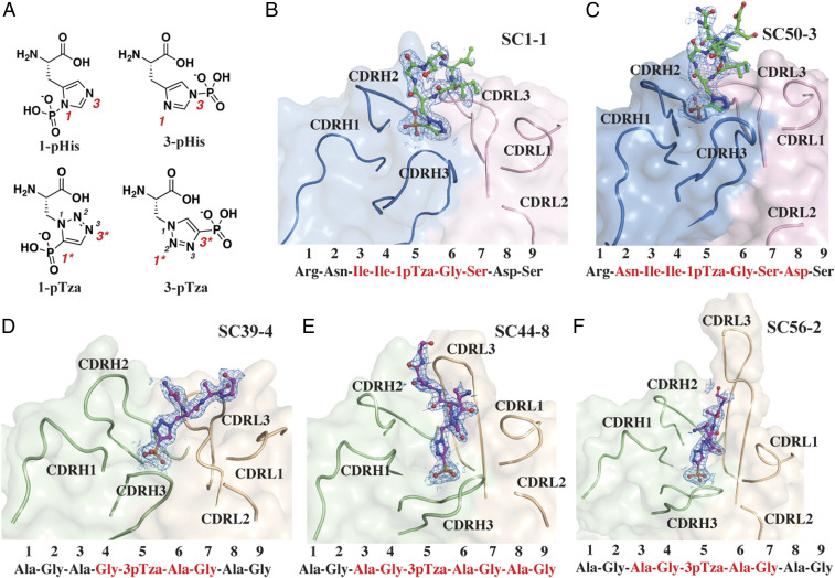
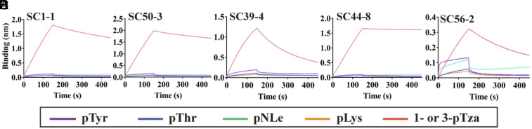
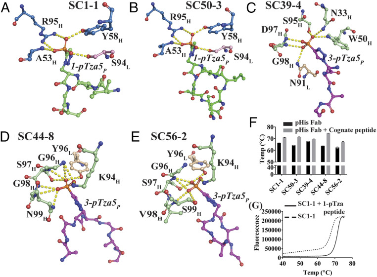
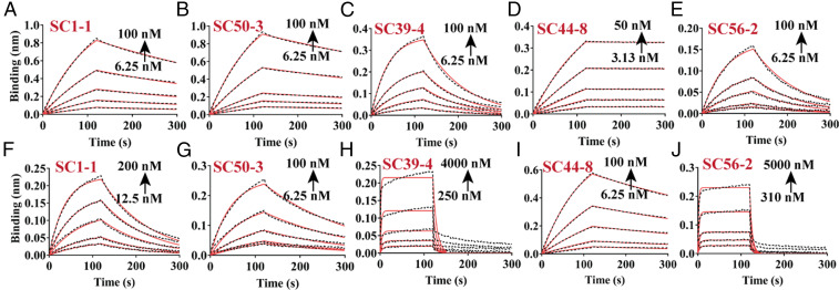
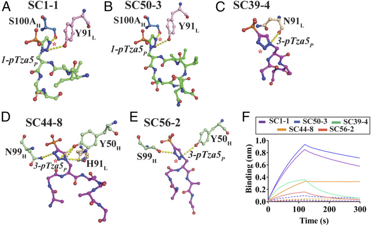
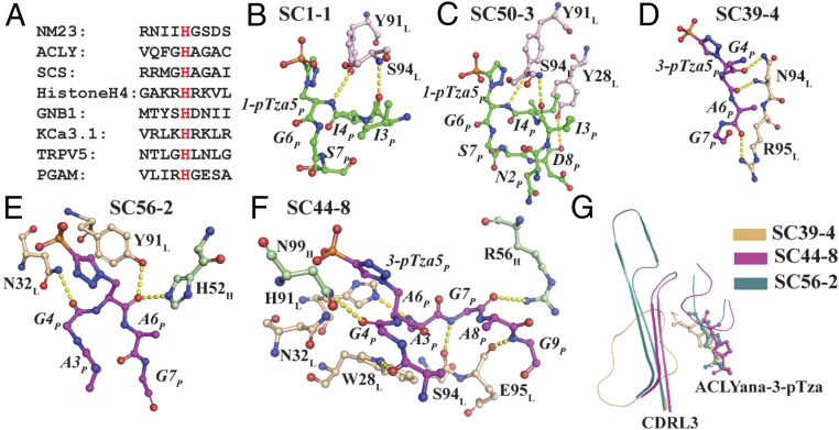
In 2015, monoclonal antibodies (mAbs) that selectively recognize the 1-pHis or 3-pHis isoforms of phosphohistidine were developed by immunizing rabbits with degenerate Ala/Gly peptides containing the nonhydrolyzable phosphohistidine (pHis) analog- phosphotriazolylalanine (pTza). Here, we report structures of five rabbit mAbs bound to cognate pTza peptides: SC1-1 and SC50-3 that recognize 1-pHis, and their 3-pHis-specific counterparts, SC39-4, SC44-8, and SC56-2. These cocrystal structures provide insights into the binding modes of the pTza phosphate group that are distinct for the 1- and 3-pHis mAbs with the selectivity arising from specific contacts with the phosphate group and triazolyl ring. The mode of phosphate recognition in the 3-pHis mAbs recapitulates the Walker A motif, as present in kinases. The complementarity-determining regions (CDRs) of four of the Fabs interact with the peptide backbone rather than peptide side chains, thus conferring sequence independence, whereas SC44-8 shows a proclivity for binding a GpHAGA motif mediated by a sterically complementary CDRL3 loop. Specific hydrogen bonding with the triazolyl ring precludes recognition of pTyr and other phosphoamino acids by these mAbs. Kinetic binding experiments reveal that the affinity of pHis mAbs for pHis and pTza peptides is submicromolar. Bound pHis mAbs also shield the pHis peptides from rapid dephosphorylation. The epitope-paratope interactions illustrate how these anti-pHis antibodies are useful for a wide range of research techniques and this structural information can be utilized to improve the specificity and affinity of these antibodies toward a variety of pHis substrates to understand the role of histidine phosphorylation in healthy and diseased states.
2015年,通过用含有不可水解的磷酸组氨酸(pHis)类似物——磷酸三唑基丙氨酸(pTza)的简并丙氨酸/甘氨酸肽免疫兔子,开发出了选择性识别磷酸组氨酸1 - pHis或3 - pHis异构体的单克隆抗体(mAb)。在此,我们报告了五种与同源pTza肽结合的兔单克隆抗体的结构:识别1 - pHis的SC1 - 1和SC50 - 3,以及它们识别3 - pHis的对应抗体SC39 - 4、SC44 - 8和SC56 - 2。这些共晶体结构揭示了pTza磷酸基团的结合模式,1 - pHis和3 - pHis单克隆抗体的结合模式不同,选择性源于与磷酸基团和三唑环的特定接触。3 - pHis单克隆抗体中磷酸的识别模式重现了激酶中存在的沃克A基序。四种Fab的互补决定区(CDR)与肽主链而非肽侧链相互作用,从而赋予序列独立性,而SC44 - 8表现出结合由空间互补的CDRL3环介导的GpHAGA基序的倾向。与三唑环的特异性氢键作用排除了这些单克隆抗体对pTyr和其他磷酸氨基酸的识别。动力学结合实验表明,pHis单克隆抗体对pHis和pTza肽的亲和力为亚微摩尔级。结合的pHis单克隆抗体还能保护pHis肽不被快速去磷酸化。抗原表位 - 抗体互补位相互作用说明了这些抗pHis抗体如何可用于广泛的研究技术,并且这一结构信息可用于提高这些抗体对各种pHis底物的特异性和亲和力,以了解组氨酸磷酸化在健康和疾病状态中的作用。