• 文献检索
  • 文档翻译
  • 深度研究
  • 学术资讯
  • Suppr Zotero 插件Zotero 插件
  • 邀请有礼
  • 套餐&价格
  • 历史记录
应用&插件
Suppr Zotero 插件Zotero 插件浏览器插件Mac 客户端Windows 客户端微信小程序
定价
高级版会员购买积分包购买API积分包
服务
文献检索文档翻译深度研究API 文档MCP 服务
关于我们
关于 Suppr公司介绍联系我们用户协议隐私条款
关注我们

Suppr 超能文献

核心技术专利:CN118964589B侵权必究
粤ICP备2023148730 号-1Suppr @ 2026

文献检索

告别复杂PubMed语法,用中文像聊天一样搜索,搜遍4000万医学文献。AI智能推荐,让科研检索更轻松。

立即免费搜索

文件翻译

保留排版,准确专业,支持PDF/Word/PPT等文件格式,支持 12+语言互译。

免费翻译文档

深度研究

AI帮你快速写综述,25分钟生成高质量综述,智能提取关键信息,辅助科研写作。

立即免费体验

YAP激活及其在胆道闭锁患者和小鼠模型中的意义

YAP Activation and Implications in Patients and a Mouse Model of Biliary Atresia.

作者信息

Zheng Chao, Luo Jiaqian, Yang Yifan, Dong Rui, Yu Fa-Xing, Zheng Shan

机构信息

Department of Pediatric Surgery, Children's Hospital of Fudan University, Shanghai, China.

Institute of Pediatrics, Children's Hospital of Fudan University and Institutes of Biomedical Sciences, Shanghai Medical College, Fudan University, Shanghai, China.

出版信息

Front Pediatr. 2021 Jan 21;8:618226. doi: 10.3389/fped.2020.618226. eCollection 2020.

DOI:10.3389/fped.2020.618226
PMID:33553074
原文链接:https://pmc.ncbi.nlm.nih.gov/articles/PMC7859521/
Abstract

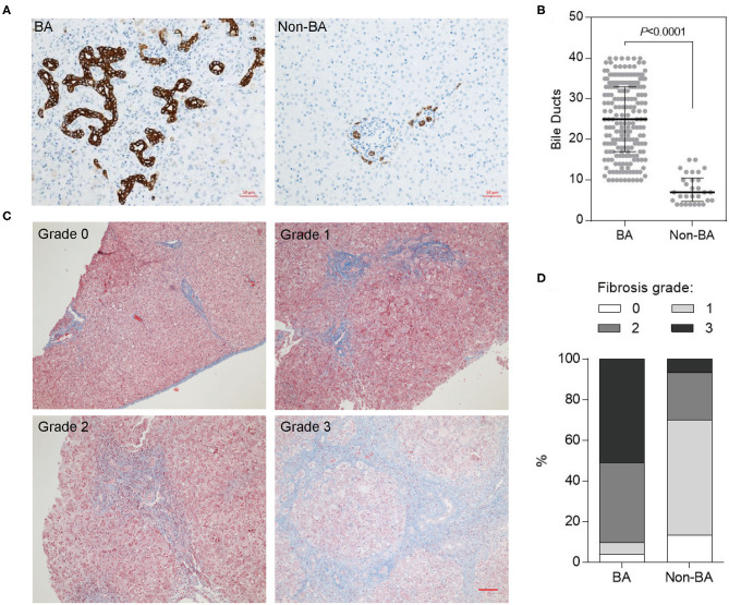
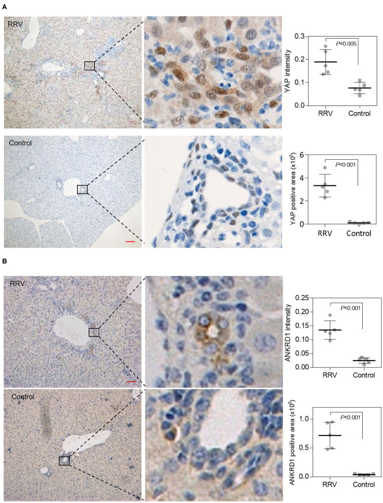
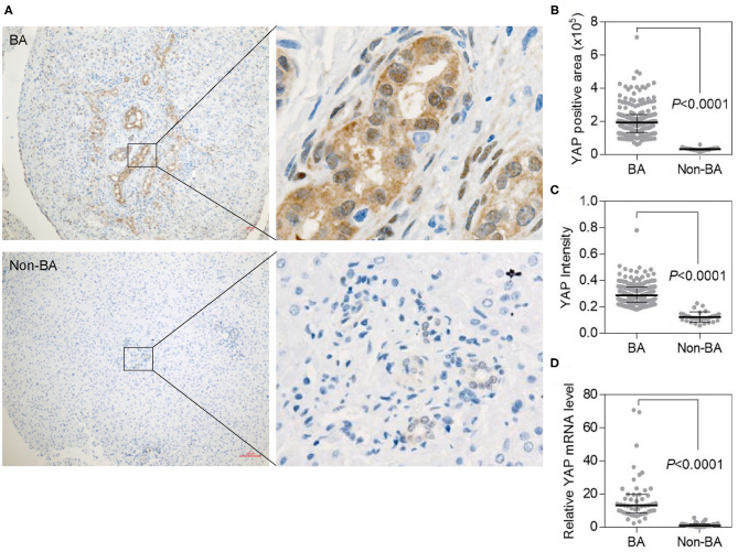
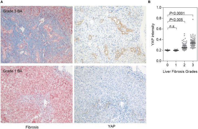
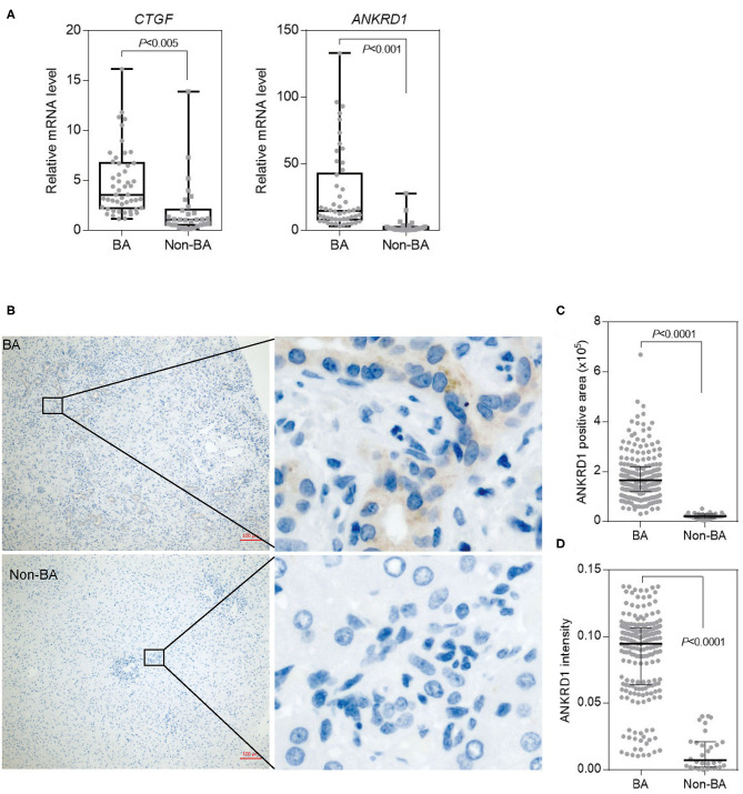
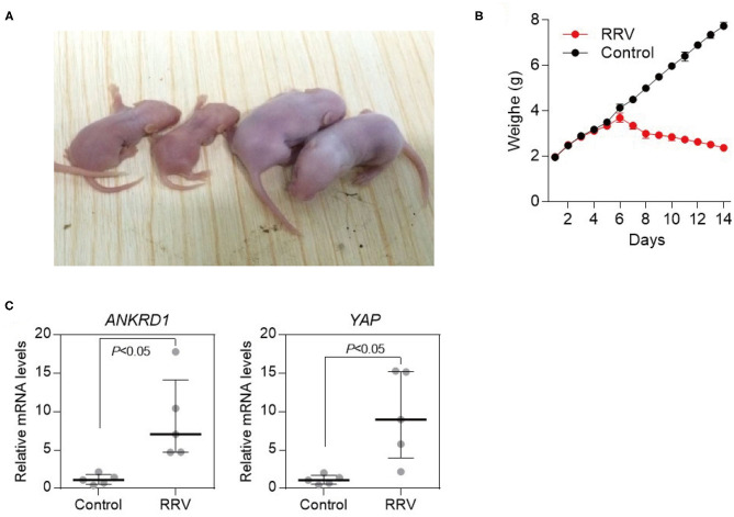
Biliary atresia (BA), an inflammatory destruction of the bile ducts, leads to liver fibrosis in infants and accounts for half of cases undergoing pediatric liver transplantation. Yes-associated protein (YAP), an effector of the Hippo signaling pathway, is critical in maintaining identities of bile ductal cells. Here, we evaluated the expression of YAP and YAP target genes in BA livers and a rhesus rotavirus (RRV)-induced BA mice model. Liver specimens collected from 200 BA patients were compared with those of 30 non-BA patients. Model mice liver tissues were also collected. The expression of YAP and YAP target genes were measured by transfection, RNA-seq, immunohistochemistry, immunoblot, and quantitative PCR. Masson's trichrome staining and the Biliary Atresia Research Consortium (BARC) system were utilized to score liver fibrosis status. The expression of YAP is elevated and positively correlated with fibrosis in BA livers. Moreover, , which is identified as the target gene of YAP, is also highly expressed in BA livers. Consistent with clinical data, YAP and ANKRD1 are significantly upregulated in RRV-induced BA mouse model. YAP expression is closely correlated with the bile duct hyperplasia and liver fibrosis, and may serve as an indicator for liver fibrosis and BA progression. This study indicates an involvement of the Hippo signaling pathway in the development of BA, and the YAP induced ANKRD1 expression may also be related to bile duct hyperplasia in BA. This provides a new direction for more in-depth exploration of the etiology and pathogenesis of biliary atresia.

摘要

胆道闭锁(BA)是一种胆管的炎性破坏疾病,可导致婴儿肝纤维化,且占小儿肝移植病例的一半。Yes相关蛋白(YAP)是Hippo信号通路的效应器,对维持胆管细胞的特性至关重要。在此,我们评估了YAP及其靶基因在BA肝脏和恒河猴轮状病毒(RRV)诱导的BA小鼠模型中的表达。将收集的200例BA患者的肝脏标本与30例非BA患者的标本进行比较。同时也收集模型小鼠的肝脏组织。通过转染、RNA测序、免疫组织化学、免疫印迹和定量PCR检测YAP及其靶基因的表达。采用Masson三色染色法和胆道闭锁研究联盟(BARC)系统对肝纤维化状态进行评分。YAP在BA肝脏中的表达升高,且与纤维化呈正相关。此外,被确定为YAP靶基因的ANKRD1在BA肝脏中也高表达。与临床数据一致,YAP和ANKRD1在RRV诱导的BA小鼠模型中显著上调。YAP表达与胆管增生和肝纤维化密切相关,可能作为肝纤维化和BA进展的一个指标。本研究表明Hippo信号通路参与了BA的发生发展,YAP诱导的ANKRD1表达也可能与BA中的胆管增生有关。这为更深入探究胆道闭锁的病因和发病机制提供了新方向。

https://cdn.ncbi.nlm.nih.gov/pmc/blobs/3074/7859521/389f319cbf4f/fped-08-618226-g0006.jpg
https://cdn.ncbi.nlm.nih.gov/pmc/blobs/3074/7859521/77ca78adf749/fped-08-618226-g0001.jpg
https://cdn.ncbi.nlm.nih.gov/pmc/blobs/3074/7859521/5de5c8c00c74/fped-08-618226-g0002.jpg
https://cdn.ncbi.nlm.nih.gov/pmc/blobs/3074/7859521/956546b6ce1e/fped-08-618226-g0003.jpg
https://cdn.ncbi.nlm.nih.gov/pmc/blobs/3074/7859521/224eb2a0a02b/fped-08-618226-g0004.jpg
https://cdn.ncbi.nlm.nih.gov/pmc/blobs/3074/7859521/044a28339a72/fped-08-618226-g0005.jpg
https://cdn.ncbi.nlm.nih.gov/pmc/blobs/3074/7859521/389f319cbf4f/fped-08-618226-g0006.jpg

相似文献

1
YAP Activation and Implications in Patients and a Mouse Model of Biliary Atresia.YAP激活及其在胆道闭锁患者和小鼠模型中的意义
Front Pediatr. 2021 Jan 21;8:618226. doi: 10.3389/fped.2020.618226. eCollection 2020.
2
RRV-induced biliary atresia in neonatal mice involves CD8 + T lymphocyte killer cells and the Notch signaling pathway.RRV 诱导的新生小鼠胆道闭锁涉及 CD8+T 淋巴细胞杀伤细胞和 Notch 信号通路。
Genes Genomics. 2021 Nov;43(11):1289-1299. doi: 10.1007/s13258-021-01153-5. Epub 2021 Aug 19.
3
The use of Yes-associated protein expression in the diagnosis of persistent neonatal cholestatic liver disease.Yes 相关蛋白表达在新生儿胆汁淤积性肝病持续期诊断中的应用。
Hum Pathol. 2014 May;45(5):1057-64. doi: 10.1016/j.humpath.2014.01.002. Epub 2014 Jan 23.
4
Liver fibrosis during the development of biliary atresia: Proof of principle in the murine model.胆道闭锁发展过程中的肝纤维化:小鼠模型中的原理验证
J Pediatr Surg. 2015 Aug;50(8):1304-9. doi: 10.1016/j.jpedsurg.2014.12.027. Epub 2015 Jan 23.
5
Gene expression profile of TLR7 signaling pathway in the liver of rhesus rotavirus-induced murine biliary atresia.TLR7 信号通路在恒河猴轮状病毒诱导的胆道闭锁小鼠肝脏中的基因表达谱。
Biochem Biophys Res Commun. 2018 Sep 3;503(1):291-296. doi: 10.1016/j.bbrc.2018.06.018. Epub 2018 Jun 22.
6
Regulation of epithelial injury and bile duct obstruction by NLRP3, IL-1R1 in experimental biliary atresia.NLRP3、IL-1R1 在实验性胆道闭锁中对胆管上皮损伤和胆管阻塞的调控作用。
J Hepatol. 2018 Nov;69(5):1136-1144. doi: 10.1016/j.jhep.2018.05.038. Epub 2018 Jun 8.
7
Regulatory T cells inhibit Th1 cell-mediated bile duct injury in murine biliary atresia.调节性 T 细胞抑制鼠先天性胆道闭锁中 Th1 细胞介导的胆管损伤。
J Hepatol. 2013 Oct;59(4):790-6. doi: 10.1016/j.jhep.2013.05.010. Epub 2013 May 14.
8
Interleukin 17, Produced by γδ T Cells, Contributes to Hepatic Inflammation in a Mouse Model of Biliary Atresia and Is Increased in Livers of Patients.γδ T 细胞产生的白细胞介素 17 促进胆道闭锁小鼠模型的肝炎症反应,并在患者肝脏中增加。
Gastroenterology. 2016 Jan;150(1):229-241.e5. doi: 10.1053/j.gastro.2015.09.008. Epub 2015 Sep 25.
9
Abnormal activation of OPN inflammation pathway in livers of children with biliary atresia and relationship to hepatic fibrosis.胆道闭锁患儿肝脏中骨桥蛋白炎症通路的异常激活及其与肝纤维化的关系。
Eur J Pediatr Surg. 2008 Aug;18(4):224-9. doi: 10.1055/s-2008-1038483. Epub 2008 Aug 14.
10
Rhesus rotavirus VP4 sequence-specific activation of mononuclear cells is associated with cholangiopathy in murine biliary atresia.恒河猴轮状病毒VP4对单核细胞的序列特异性激活与小鼠胆道闭锁中的胆管病相关。
Am J Physiol Gastrointest Liver Physiol. 2015 Sep 15;309(6):G466-74. doi: 10.1152/ajpgi.00079.2015. Epub 2015 Jul 23.

引用本文的文献

1
Single-cell transcription reveals hepatocyte-to-cholangiocyte reprogramming and biliary gene profile in biliary atresia.单细胞转录揭示了胆道闭锁中肝细胞向胆管细胞的重编程及胆管基因谱。
Hepatol Commun. 2025 May 6;9(5). doi: 10.1097/HC9.0000000000000710. eCollection 2025 May 1.
2
The Hippo signalling pathway and its impact on eye diseases.Hippo 信号通路及其对眼部疾病的影响。
J Cell Mol Med. 2024 Apr;28(8):e18300. doi: 10.1111/jcmm.18300.
3
Cellular and Molecular Mechanisms of Toxic Liver Fibrosis in Rats Depending on the Stages of Its Development.

本文引用的文献

1
Blockade of YAP alleviates hepatic fibrosis through accelerating apoptosis and reversion of activated hepatic stellate cells.阻断 YAP 可通过加速细胞凋亡和活化的肝星状细胞的逆转来减轻肝纤维化。
Mol Immunol. 2019 Mar;107:29-40. doi: 10.1016/j.molimm.2019.01.004. Epub 2019 Jan 11.
2
Proliferation of hepatic stellate cells, mediated by YAP and TAZ, contributes to liver repair and regeneration after liver ischemia-reperfusion injury.YAP 和 TAZ 介导的肝星状细胞增殖有助于肝缺血再灌注损伤后的肝修复和再生。
Am J Physiol Gastrointest Liver Physiol. 2018 Apr 1;314(4):G471-G482. doi: 10.1152/ajpgi.00153.2017. Epub 2018 Jan 11.
3
大鼠中毒性肝纤维化根据其发展阶段的细胞和分子机制
Sovrem Tekhnologii Med. 2023;15(4):50-63. doi: 10.17691/stm2023.15.4.05. Epub 2023 Jul 28.
4
literammp-9 mRNA Expression and Bridging Fibrosis Progression in Toxic Liver Injury.肝再生增强因子-9信使核糖核酸在中毒性肝损伤中的表达及与桥接纤维化进展的关系
Acta Naturae. 2023 Apr-Jun;15(2):50-58. doi: 10.32607/actanaturae.17856.
5
Role of YAP1 Signaling in Biliary Development, Repair, and Disease.YAP1 信号在胆管发育、修复和疾病中的作用。
Semin Liver Dis. 2022 Feb;42(1):17-33. doi: 10.1055/s-0041-1742277. Epub 2022 Jan 24.
Intrahepatic Biliary Metastasis of Colonic Adenocarcinoma: A Case Report With Immunohistochemical Analysis.
结肠腺癌肝内胆管转移:一例免疫组织化学分析病例报告
World J Oncol. 2017 Jun;8(3):86-91. doi: 10.14740/wjon1037w. Epub 2017 Jun 9.
4
The Hippo signaling functions through the Notch signaling to regulate intrahepatic bile duct development in mammals.河马信号通路通过Notch信号通路发挥作用,以调节哺乳动物肝内胆管的发育。
Lab Invest. 2017 Jul;97(7):843-853. doi: 10.1038/labinvest.2017.29. Epub 2017 Jun 5.
5
Altered Expression of Hippo Signaling Pathway Molecules in Intrahepatic Cholangiocarcinoma.肝内胆管癌中Hippo信号通路分子的表达改变
Oncology. 2017;93(1):67-74. doi: 10.1159/000463390. Epub 2017 Apr 28.
6
Forkhead box A3 attenuated the progression of fibrosis in a rat model of biliary atresia.叉头框A3减轻了胆道闭锁大鼠模型中的纤维化进程。
Cell Death Dis. 2017 Mar 30;8(3):e2719. doi: 10.1038/cddis.2017.99.
7
Flow-dependent YAP/TAZ activities regulate endothelial phenotypes and atherosclerosis.血流依赖性YAP/TAZ活性调节内皮细胞表型和动脉粥样硬化。
Proc Natl Acad Sci U S A. 2016 Oct 11;113(41):11525-11530. doi: 10.1073/pnas.1613121113. Epub 2016 Sep 26.
8
Ankyrin Repeat Domain 1 is Up-regulated During Hepatitis C Virus Infection and Regulates Hepatitis C Virus Entry.锚蛋白重复结构域1在丙型肝炎病毒感染期间上调并调节丙型肝炎病毒进入。
Sci Rep. 2016 Feb 10;6:20819. doi: 10.1038/srep20819.
9
Hippo Pathway in Organ Size Control, Tissue Homeostasis, and Cancer.器官大小调控、组织稳态及癌症中的河马信号通路
Cell. 2015 Nov 5;163(4):811-28. doi: 10.1016/j.cell.2015.10.044.
10
The Hippo pathway effector YAP controls mouse hepatic stellate cell activation.Hippo 通路效应物 YAP 控制小鼠肝星状细胞的激活。
J Hepatol. 2015 Sep;63(3):679-88. doi: 10.1016/j.jhep.2015.04.011. Epub 2015 Apr 20.