Kawashima Keisuke, Watanabe Naoki, Tawada Sho, Adachi Takahito, Yamada Makoto, Kitoh Yusuke, Takeuchi Tamotsu, Tanaka Takuji
Department of Pathology and Translational Research, Gifu University School of Mediine, 1-1 Yanagido, Gifu City, Gifu 501-1194, Japan.
Department of Diagnostic Pathology (DDP) & Research Center of Diagnostic Pathology (RC-DiP), Gifu Municipal Hospital, 7-1 Kashima-cho, Gifu City, Gifu 500-8513, Japan.
World J Oncol. 2017 Jun;8(3):86-91. doi: 10.14740/wjon1037w. Epub 2017 Jun 9.
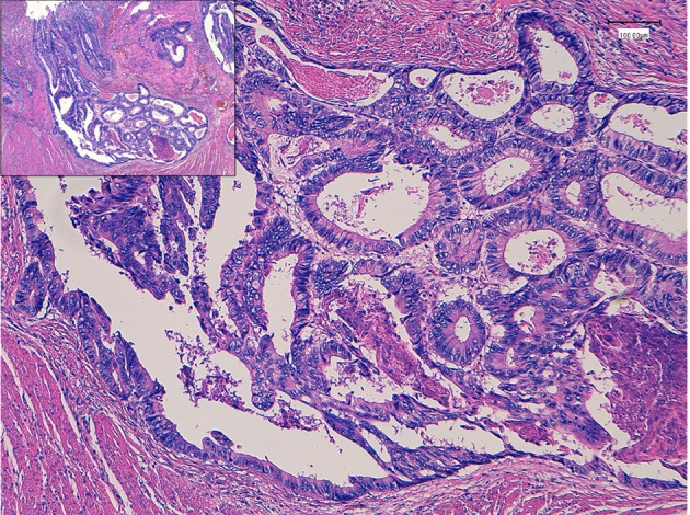
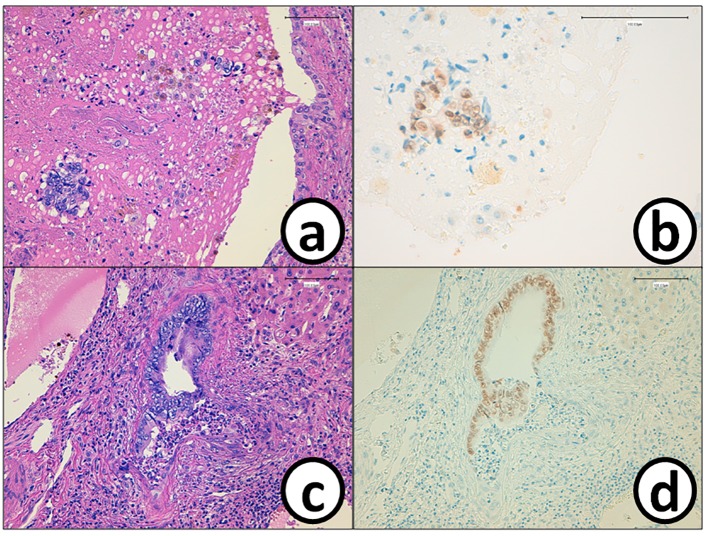
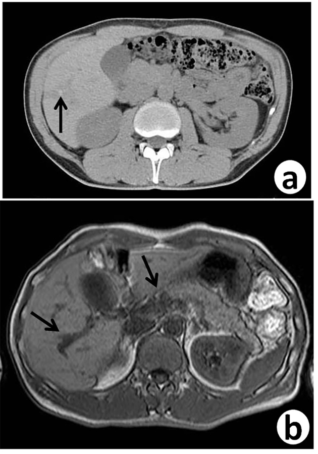
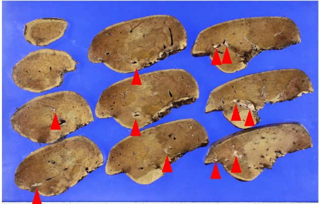
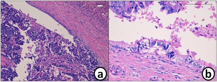
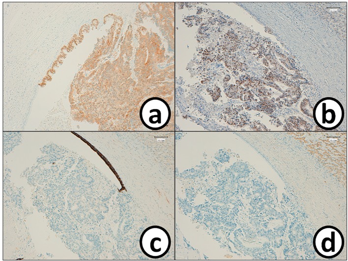
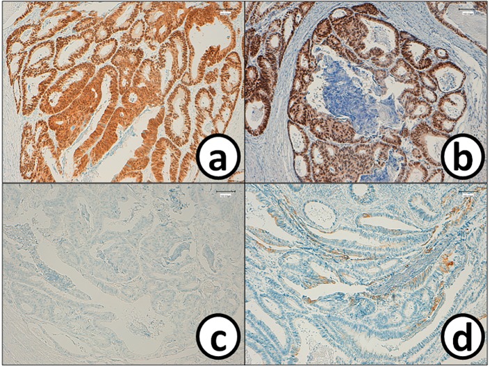
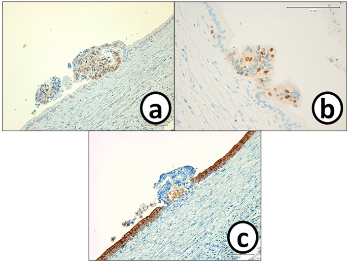
Although intrabiliary metastasis of carcinoma in the liver is unusual, intraductal and/or intraepithelial spread of cancer cells along intrahepatic bile ducts is now well recognized as hepatic metastasis. However, several clinical and laboratory findings, including images, lead us to differentially diagnose from primary intrahepatic cholangiocarcinoma. We report here on a case of colonic adenocarcinoma that metastasized to the liver with spread along with the intrahepatic bile duct of S5/6 area. The patient was a 51-year-old man and clinically diagnosed liver metastasis of sigmoid colon cancer (tub2, pMP, ly1, v0, n0), which was diagnosed and treated by sigmoidectomy 7 years ago. The right hepatic lobectomy was performed in March 2016 and histopathological examination revealed that moderately differentiated adenocarcinoma proliferated along the epithelium of intrahepatic bile ducts. Immunohistochemistry (IHC) showed that cancer cells in the intrahepatic bile ducts were positive for CK20, CDX2, CK17 and CK19, but negative for CK7, MUC-5AC, MUC-2 and CA19-9. The findings were almost the same as those of the sigmoid colon cancer removed in July 2009. We finally diagnosed the liver tumor as intrahepatic biliary metastasis of sigmoid colon cancer. Patients with liver metastasis of cancer are hard to be detected biliary invasion and spread on diagnostic image examination. Knowledge of distinctive morphological and IHC features can help to accurately diagnose this rare intrahepatic biliary metastasis of colonic cancer in routine pathological diagnostic procedures.
尽管肝癌的肝内胆管转移并不常见,但癌细胞沿肝内胆管的导管内和/或上皮内扩散现在已被公认为肝转移。然而,包括影像学在内的一些临床和实验室检查结果,使我们需要与原发性肝内胆管癌进行鉴别诊断。我们在此报告一例结肠腺癌肝转移病例,癌细胞沿S5/6区肝内胆管扩散。患者为51岁男性,临床诊断为乙状结肠癌肝转移(tub2,pMP,ly1,v0,n0),7年前已行乙状结肠切除术。2016年3月进行了右肝叶切除术,组织病理学检查显示中分化腺癌沿肝内胆管上皮增生。免疫组化(IHC)显示肝内胆管癌细胞CK20、CDX2、CK17和CK19呈阳性,但CK7、MUC-5AC、MUC-2和CA19-9呈阴性。这些结果与2009年7月切除的乙状结肠癌几乎相同。我们最终诊断肝脏肿瘤为乙状结肠癌肝内胆管转移。癌症肝转移患者在诊断性影像检查中很难检测到胆管侵犯和扩散。了解独特的形态学和免疫组化特征有助于在常规病理诊断过程中准确诊断这种罕见的结肠癌肝内胆管转移。