Department of Nanomedicine & Drug Targeting, Groningen Research Institute of Pharmacy, University of Groningen, A. Deusinglaan 1, 9713 AV, Groningen, The Netherlands.
Department of Pharmaceutical Technology and Biopharmacy, Groningen Research Institute of Pharmacy, University of Groningen, A. Deusinglaan 1, 9713 AV, Groningen, The Netherlands.
Arch Toxicol. 2021 Apr;95(4):1267-1285. doi: 10.1007/s00204-021-02992-7. Epub 2021 Feb 8.
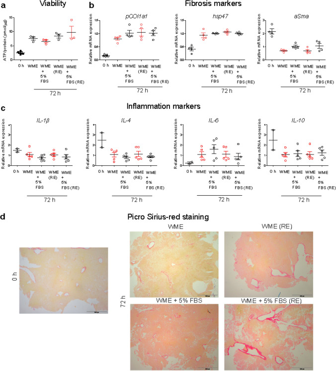
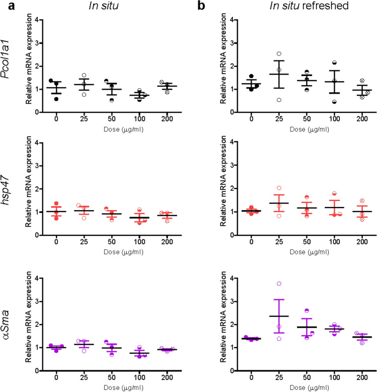
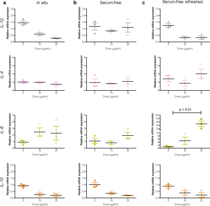
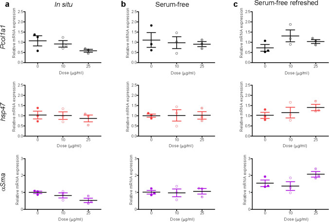
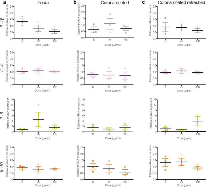
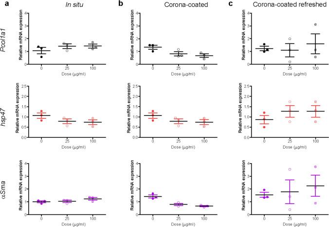
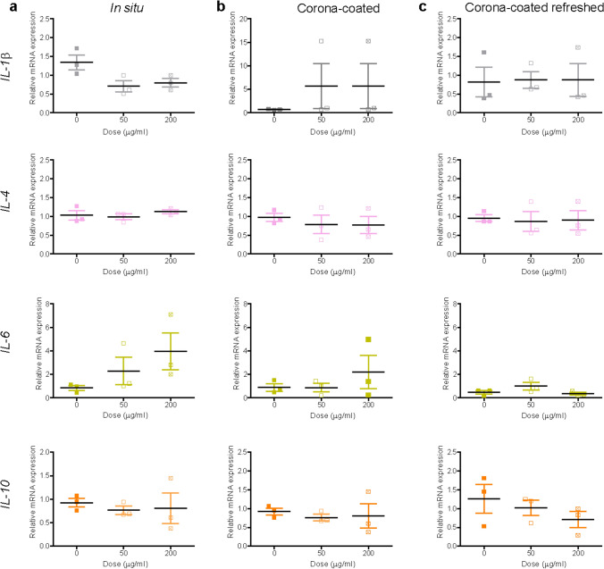
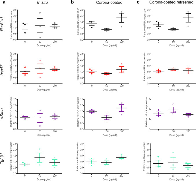
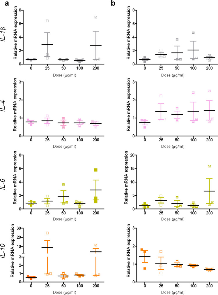
Chronic exposure and accumulation of persistent nanomaterials by cells have led to safety concerns on potential long-term effects induced by nanoparticles, including chronic inflammation and fibrosis. With this in mind, we used murine precision-cut liver tissue slices to test potential induction of inflammation and onset of fibrosis upon 72 h exposure to different nanomaterials (0-200 µg/ml). Tissue slices were chosen as an advanced ex vivo 3D model to better resemble the complexity of the in vivo tissue environment, with a focus on the liver where most nanomaterials accumulate. Effects on the onset of fibrosis and inflammation were investigated, with particular care in optimizing nanoparticle exposure conditions to tissue. Thus, we compared the effects induced on slices exposed to nanoparticles in the presence of excess free proteins (in situ), or after corona isolation. Slices exposed to daily-refreshed nanoparticle dispersions were used to test additional effects due to ageing of the dispersions. Exposure to amino-modified polystyrene nanoparticles in serum-free conditions led to strong inflammation, with stronger effects with daily-refreshed dispersions. Instead, no inflammation was observed when slices were exposed to the same nanoparticles in medium supplemented with serum to allow corona formation. Similarly, no clear signs of inflammation nor of onset of fibrosis were detected after exposure to silica, titania or carboxylated polystyrene in all conditions tested. Overall, these results show that liver slices can be used to test nanoparticle-induced inflammation in real tissue, and that the exposure conditions and ageing of the dispersions can strongly affect tissue responses to nanoparticles.
慢性暴露和细胞内持久性纳米材料的积累导致了人们对纳米颗粒潜在长期影响的安全担忧,包括慢性炎症和纤维化。考虑到这一点,我们使用鼠精密切割肝组织切片来测试不同纳米材料(0-200μg/ml)72 小时暴露后潜在的炎症诱导和纤维化发病。组织切片被选为一种先进的离体 3D 模型,以更好地模拟体内组织环境的复杂性,重点是纳米材料最容易积累的肝脏。研究了对纤维化和炎症发病的影响,并特别注意优化纳米颗粒暴露条件对组织的影响。因此,我们比较了在存在过量游离蛋白的情况下(原位)或在冠状隔离后暴露于纳米颗粒的切片上诱导的效果。每天更新纳米颗粒分散体的切片用于测试由于分散体老化而引起的额外效果。在无血清条件下暴露于氨基修饰的聚苯乙烯纳米颗粒会导致强烈的炎症,而每天更新的分散体则会产生更强的效果。相反,当在含有血清的培养基中暴露于相同的纳米颗粒以允许形成冠状物时,未观察到切片有炎症。同样,在所有测试条件下,暴露于二氧化硅、二氧化钛或羧化聚苯乙烯后,未检测到明显的炎症或纤维化发病迹象。总的来说,这些结果表明,肝切片可用于在真实组织中测试纳米颗粒诱导的炎症,并且分散体的暴露条件和老化会强烈影响组织对纳米颗粒的反应。