• 文献检索
  • 文档翻译
  • 深度研究
  • 学术资讯
  • Suppr Zotero 插件Zotero 插件
  • 邀请有礼
  • 套餐&价格
  • 历史记录
应用&插件
Suppr Zotero 插件Zotero 插件浏览器插件Mac 客户端Windows 客户端微信小程序
定价
高级版会员购买积分包购买API积分包
服务
文献检索文档翻译深度研究API 文档MCP 服务
关于我们
关于 Suppr公司介绍联系我们用户协议隐私条款
关注我们

Suppr 超能文献

核心技术专利:CN118964589B侵权必究
粤ICP备2023148730 号-1Suppr @ 2026

文献检索

告别复杂PubMed语法,用中文像聊天一样搜索,搜遍4000万医学文献。AI智能推荐,让科研检索更轻松。

立即免费搜索

文件翻译

保留排版,准确专业,支持PDF/Word/PPT等文件格式,支持 12+语言互译。

免费翻译文档

深度研究

AI帮你快速写综述,25分钟生成高质量综述,智能提取关键信息,辅助科研写作。

立即免费体验

玻璃体后炎症的自动量化:光学相干断层扫描扫描数量要求。

Automated quantification of posterior vitreous inflammation: optical coherence tomography scan number requirements.

机构信息

Department of Ophthalmology, University Hospital Bonn, 53127, Bonn, Germany.

Division of Optometry and Visual Science, City, University of London, London, UK.

出版信息

Sci Rep. 2021 Feb 8;11(1):3271. doi: 10.1038/s41598-021-82786-0.

DOI:10.1038/s41598-021-82786-0
PMID:33558619
原文链接:https://pmc.ncbi.nlm.nih.gov/articles/PMC7870671/
Abstract

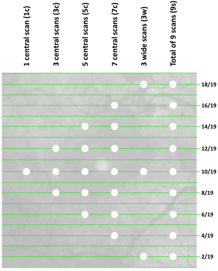
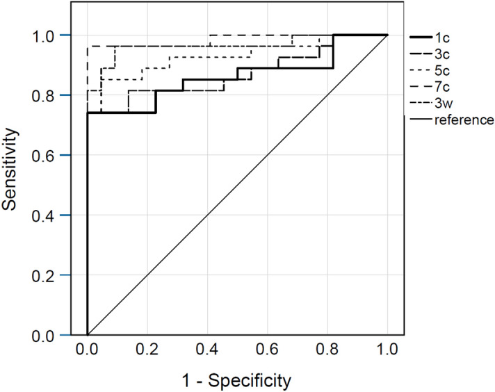
Quantifying intraocular inflammation is crucial in managing uveitis patients. We assessed the minimum B-scan density for reliable automated vitreous intensity (VI) assessment, using a novel approach based on optical coherence tomography (OCT). OCT volume scans centered on the macula were retrospectively collected in patients with uveitis. Nine B-scans per volume scan at fixed locations were automatically analyzed. The following B-scan selections were compared against the average score of 9 B-scans per volume scan as a reference standard: 1/3/5/7 central scans (1c/3c/5c/7c), 3 widely distributed scans (3w). Image data of 49 patients (31 females) were included. The median VI was 0.029 (IQR: 0.032). The intra-class-correlation coefficient of the VI across the 9 B-scans was 0.923. The median difference from the reference standard ranged between 0.001 (7c) and 0.006 (1c). It was significantly lower for scan selection 3w than 5c, p(adjusted) = 0.022, and lower for selection 7c than 3w, p(adjusted) = 0.003. The scan selections 7c and 3w showed the two highest areas under the receiver operating curve (0.985 and 0.965, respectively). Three widely distributed B-scans are sufficient to quantify VI reliably. Highest reliability was achieved using 7 central B-scans. Automated quantification of VI in uveitis is reliable and requires only few OCT B-scans.

摘要

量化眼内炎症对于管理葡萄膜炎患者至关重要。我们使用基于光学相干断层扫描(OCT)的新方法评估了可靠的自动玻璃体强度(VI)评估的最小 B 扫描密度。回顾性收集了葡萄膜炎患者的黄斑中心 OCT 容积扫描。在固定位置自动分析每个容积扫描的 9 个 B 扫描。以下 B 扫描选择与每个容积扫描的 9 个 B 扫描的平均得分(参考标准)进行比较:1/3/5/7 个中央扫描(1c/3c/5c/7c),3 个广泛分布的扫描(3w)。纳入了 49 名患者(31 名女性)的图像数据。VI 的中位数为 0.029(IQR:0.032)。9 个 B 扫描之间 VI 的组内相关系数为 0.923。与参考标准的中位数差异在 0.001(7c)至 0.006(1c)之间。与 5c 相比,3w 的扫描选择差异明显更低,p(调整)= 0.022,与 7c 相比,3w 的扫描选择差异也更低,p(调整)= 0.003。7c 和 3w 的扫描选择分别显示了最高的受试者工作特征曲线下面积(0.985 和 0.965)。3 个广泛分布的 B 扫描足以可靠地量化 VI。使用 7 个中央 B 扫描可获得最高的可靠性。自动量化葡萄膜炎中的 VI 是可靠的,仅需要少量的 OCT B 扫描。

https://cdn.ncbi.nlm.nih.gov/pmc/blobs/b1b4/7870671/0885ab3cf0f3/41598_2021_82786_Fig2_HTML.jpg
https://cdn.ncbi.nlm.nih.gov/pmc/blobs/b1b4/7870671/a37c3725b373/41598_2021_82786_Fig1_HTML.jpg
https://cdn.ncbi.nlm.nih.gov/pmc/blobs/b1b4/7870671/0885ab3cf0f3/41598_2021_82786_Fig2_HTML.jpg
https://cdn.ncbi.nlm.nih.gov/pmc/blobs/b1b4/7870671/a37c3725b373/41598_2021_82786_Fig1_HTML.jpg
https://cdn.ncbi.nlm.nih.gov/pmc/blobs/b1b4/7870671/0885ab3cf0f3/41598_2021_82786_Fig2_HTML.jpg

相似文献

1
Automated quantification of posterior vitreous inflammation: optical coherence tomography scan number requirements.玻璃体后炎症的自动量化:光学相干断层扫描扫描数量要求。
Sci Rep. 2021 Feb 8;11(1):3271. doi: 10.1038/s41598-021-82786-0.
2
Objective quantification of vitreous haze on optical coherence tomography scans: no evidence for relationship between uveitis and inflammation in multiple sclerosis.客观量化光学相干断层扫描检查中的玻璃体混浊:多发性硬化症中葡萄膜炎与炎症之间无关联证据。
Eur J Neurol. 2020 Jan;27(1):144-e3. doi: 10.1111/ene.14048. Epub 2019 Aug 18.
3
AUTOMATED QUANTIFICATION OF VITREOUS HYPERREFLECTIVE FOCI AND VITREOUS HAZE USING OPTICAL COHERENCE TOMOGRAPHY IN PATIENTS WITH UVEITIS.使用光学相干断层扫描对葡萄膜炎患者的玻璃体高反射灶和玻璃体混浊进行自动定量分析。
Retina. 2021 Nov 1;41(11):2342-2350. doi: 10.1097/IAE.0000000000003190.
4
OCT Assisted Quantification of Vitreous Inflammation in Uveitis.OCT 辅助定量评估葡萄膜炎患者玻璃体炎症反应。
Transl Vis Sci Technol. 2022 Jan 3;11(1):3. doi: 10.1167/tvst.11.1.3.
5
Spectral-domain Cirrus high-definition optical coherence tomography is better than time-domain Stratus optical coherence tomography for evaluation of macular pathologic features in uveitis.在葡萄膜炎黄斑病变特征评估方面,频域Cirrus高清光学相干断层扫描比时域Stratus光学相干断层扫描更好。
Am J Ophthalmol. 2008 Jun;145(6):1018-1022. doi: 10.1016/j.ajo.2008.01.021. Epub 2008 Mar 17.
6
Objective Quantification of Posterior Segment Inflammation: Measuring Vitreous Cells and Haze Using Optical Coherence Tomography.目的:后段炎症的定量分析——使用光学相干断层扫描测量玻璃体细胞和混浊
Am J Ophthalmol. 2023 Jan;245:134-144. doi: 10.1016/j.ajo.2022.08.025. Epub 2022 Sep 7.
7
Relationship between ultra-widefield optical coherence tomography and ophthalmoscopy for detecting posterior inflammation in posterior uveitis and panuveitis.超广角光学相干断层扫描与眼底镜在检测后葡萄膜炎和全葡萄膜炎后段炎症中的关系。
PLoS One. 2023 Feb 10;18(2):e0281714. doi: 10.1371/journal.pone.0281714. eCollection 2023.
8
Objective measurement of vitreous inflammation using optical coherence tomography.使用光学相干断层扫描技术对玻璃体炎症进行客观测量。
Ophthalmology. 2014 Sep;121(9):1706-14. doi: 10.1016/j.ophtha.2014.03.006. Epub 2014 May 15.
9
Automated Analysis of Anterior Chamber Inflammation by Spectral-Domain Optical Coherence Tomography.利用频域光学相干断层扫描对前房炎症进行自动分析。
Ophthalmology. 2015 Jul;122(7):1464-70. doi: 10.1016/j.ophtha.2015.02.032. Epub 2015 Apr 4.
10
Standard 6-mm Compared with Widefield 16.5-mm OCT for Staging of Posterior Vitreous Detachment.6 毫米标准与 16.5 毫米广角 OCT 在后脱离中的分期比较。
Ophthalmol Retina. 2020 Nov;4(11):1093-1102. doi: 10.1016/j.oret.2020.05.006. Epub 2020 May 19.

引用本文的文献

1
Artificial Intelligence in Uveitis: Innovations in Diagnosis and Therapeutic Strategies.葡萄膜炎中的人工智能:诊断与治疗策略的创新
Clin Ophthalmol. 2024 Dec 14;18:3753-3766. doi: 10.2147/OPTH.S495307. eCollection 2024.
2
Diagnosis, Classification, and Assessment of the Underlying Etiology of Uveitis by Artificial Intelligence: A Systematic Review.人工智能在葡萄膜炎潜在病因诊断、分类及评估中的应用:一项系统综述
J Clin Med. 2023 May 29;12(11):3746. doi: 10.3390/jcm12113746.

本文引用的文献

1
Objective quantification of vitreous haze on optical coherence tomography scans: no evidence for relationship between uveitis and inflammation in multiple sclerosis.客观量化光学相干断层扫描检查中的玻璃体混浊:多发性硬化症中葡萄膜炎与炎症之间无关联证据。
Eur J Neurol. 2020 Jan;27(1):144-e3. doi: 10.1111/ene.14048. Epub 2019 Aug 18.
2
Optimizing OCT acquisition parameters for assessments of vitreous haze for application in uveitis.优化 OCT 采集参数以评估用于葡萄膜炎的玻璃体混浊。
Sci Rep. 2018 Jan 26;8(1):1648. doi: 10.1038/s41598-018-20092-y.
3
MINIMAL OPTICAL COHERENCE TOMOGRAPHY B-SCAN DENSITY FOR RELIABLE DETECTION OF INTRARETINAL AND SUBRETINAL FLUID IN MACULAR DISEASES.
用于可靠检测黄斑病变中视网膜内和视网膜下液的最小光相干断层扫描 B 扫描密度。
Retina. 2019 Jan;39(1):150-156. doi: 10.1097/IAE.0000000000001918.
4
Biomarkers and Surrogate Endpoints in Uveitis: The Impact of Quantitative Imaging.葡萄膜炎中的生物标志物和替代终点:定量成像的影响
Invest Ophthalmol Vis Sci. 2017 May 1;58(6):BIO131-BIO140. doi: 10.1167/iovs.17-21788.
5
Impact of optical coherence tomography scanning density on quantitative analyses in neovascular age-related macular degeneration.光学相干断层扫描密度对新生血管性年龄相关性黄斑变性定量分析的影响
Eye (Lond). 2017 Jan;31(1):53-61. doi: 10.1038/eye.2016.260. Epub 2016 Dec 2.
6
A Focus on the Epidemiology of Uveitis.关注葡萄膜炎的流行病学。
Ocul Immunol Inflamm. 2018;26(1):2-16. doi: 10.1080/09273948.2016.1196713. Epub 2016 Jul 28.
7
Quantitative analysis of vitreous inflammation using optical coherence tomography in patients receiving sub-Tenon's triamcinolone acetonide for uveitic cystoid macular oedema.使用光学相干断层扫描对接受Tenon囊下曲安奈德治疗葡萄膜炎性黄斑囊样水肿患者的玻璃体炎症进行定量分析。
Br J Ophthalmol. 2017 Feb;101(2):175-179. doi: 10.1136/bjophthalmol-2015-308008. Epub 2016 May 5.
8
Evaluation of Objective Vitritis Grading Method Using Optical Coherence Tomography: Influence of Phakic Status and Previous Vitrectomy.使用光学相干断层扫描评估客观玻璃体炎分级方法:晶状体状态和既往玻璃体切除术的影响。
Am J Ophthalmol. 2016 Jan;161:172-80.e1-4. doi: 10.1016/j.ajo.2015.10.009. Epub 2015 Oct 23.
9
Automated Analysis of Vitreous Inflammation Using Spectral-Domain Optical Coherence Tomography.使用谱域光学相干断层扫描技术对玻璃体炎症进行自动分析
Transl Vis Sci Technol. 2015 Sep 16;4(5):4. doi: 10.1167/tvst.4.5.4. eCollection 2015 Sep.
10
Radial versus raster spectral-domain optical coherence tomography scan patterns for detection of macular fluid in neovascular age-related macular degeneration.用于检测新生血管性年龄相关性黄斑变性中黄斑区积液的放射状与光栅状光谱域光学相干断层扫描模式
Br J Ophthalmol. 2016 Apr;100(4):491-4. doi: 10.1136/bjophthalmol-2014-306561. Epub 2015 Aug 10.