*Institute of Vision Research, Department of Ophthalmology, Yonsei University College of Medicine, Seoul, Korea; †Corneal Dystrophy Research Institute, Yonsei University College of Medicine, Seoul, Korea; ‡Woolfson Eye Institute, Atlanta, GA; and §Saevit Eye Hospital, Goyang-Si, Gyeonggi-Do, Korea.
Cornea. 2021 Apr;40(4):519-524. doi: 10.1097/ICO.0000000000002655.
To report the outcome of unilateral small incision lenticule extraction (SMILE) in a patient with granular corneal dystrophy type 2 (GCD2).
Slit-lamp photography and Fourier domain optical coherence tomography were used to document the clinical course and appearance of the corneas in a patient with genetically determined GCD2 who underwent unilateral SMILE in the right eye.
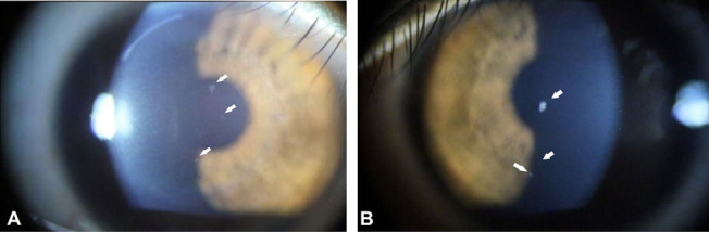
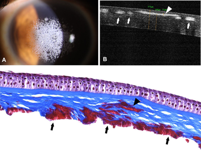
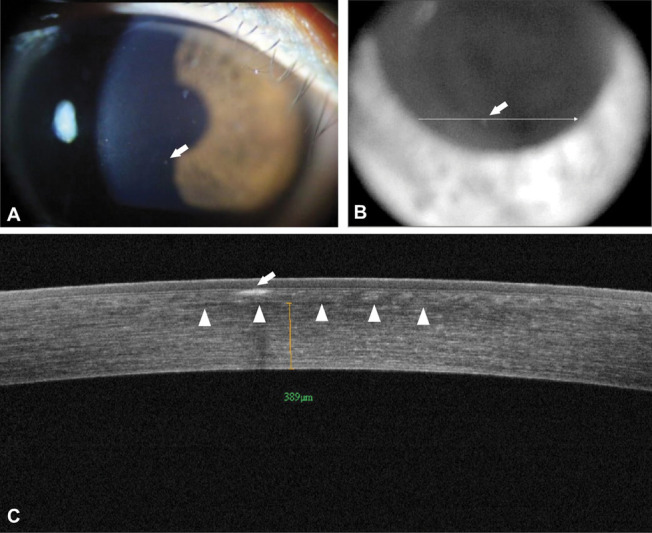
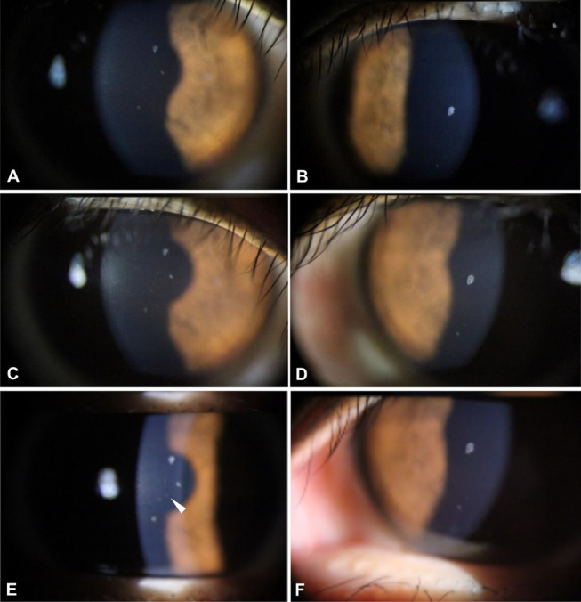
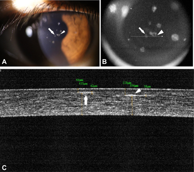
Slit-lamp examination of a 23-year-old woman revealed 2 faint opacities at the surgical interface approximately 2 months after the SMILE procedure had been performed on her right eye. Nine and 3 typical GCD2 deposits located immediately beneath the Bowman layer were observed in the right and left corneas, respectively. Over time, the deposits at the interface increased in size, density, and number in the right eye. Fourier domain optical coherence tomography performed 33 months after the SMILE procedure revealed deposits at the SMILE interface that were distinct from those located immediately beneath the Bowman layer. The severity of disease exacerbation was less in this patient than what is typically observed in others who have undergone laser-assisted in situ keratomileusis or photorefractive keratectomy.
SMILE is contraindicated in patients with GCD2, as are other corneal refractive surgical procedures. This case highlights the importance of genetic testing before the performance of refractive corneal procedures-especially for patients with corneal opacities on preoperative slit-lamp examination or a family history of corneal disease compatible with that of a corneal dystrophy.
报告 1 例颗粒状角膜营养不良 2 型(GCD2)患者行单侧小切口角膜微透镜取出术(SMILE)的结果。
使用裂隙灯摄影和傅里叶域光学相干断层扫描记录 1 例经基因诊断为 GCD2 的患者的临床病程和角膜外观,该患者在右眼行单侧 SMILE。
对 1 名 23 岁女性进行裂隙灯检查,发现右眼 SMILE 术后约 2 个月时在手术界面有 2 个隐约的混浊。右眼和左眼分别有 9 个和 3 个典型的 GCD2 沉积物位于Bowman 层下。随着时间的推移,界面处的沉积物在右眼的大小、密度和数量上均增加。SMILE 术后 33 个月行傅里叶域光学相干断层扫描显示,SMILE 界面处有沉积物,与位于 Bowman 层下的沉积物不同。与其他接受过激光辅助原位角膜磨镶术或光性角膜切削术的患者相比,该患者疾病恶化的严重程度较轻。
GCD2 患者不应行 SMILE 术或其他角膜屈光手术。本病例强调了在进行屈光性角膜手术之前进行基因检测的重要性,特别是对于术前裂隙灯检查有角膜混浊或有与角膜营养不良相符的家族史的患者。