The Russell H. Morgan Department of Radiology and Radiological Science, Johns Hopkins University School of Medicine, Baltimore, MD 21287, USA.
The Sidney Kimmel Comprehensive Cancer Center, Johns Hopkins University School of Medicine, Baltimore, MD 21231, USA.
Nanotheranostics. 2021 Jan 19;5(2):182-196. doi: 10.7150/ntno.52361. eCollection 2021.
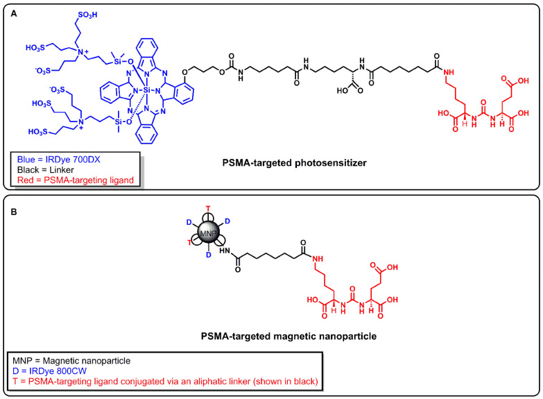
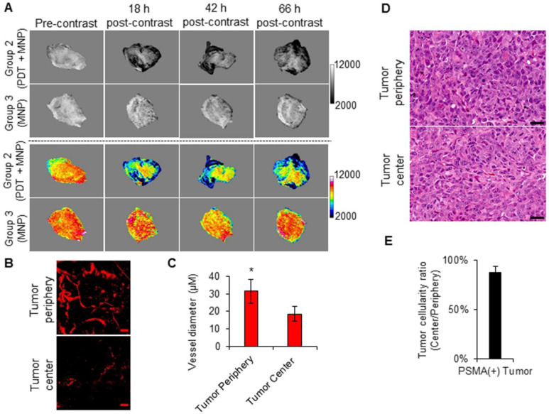
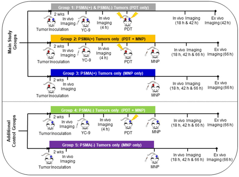
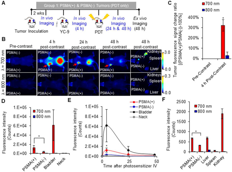
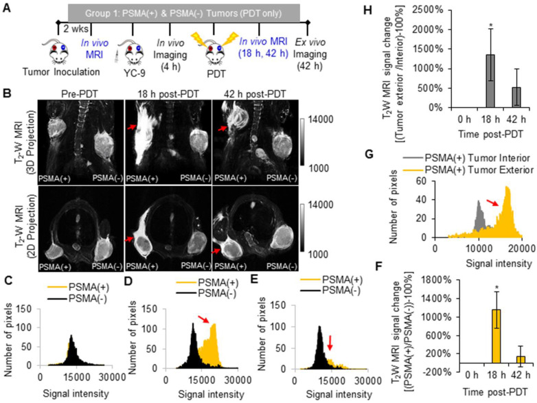
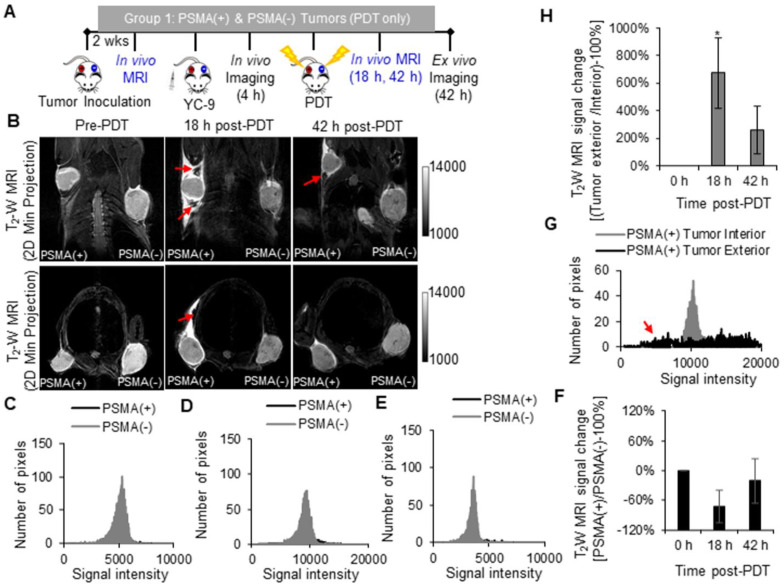
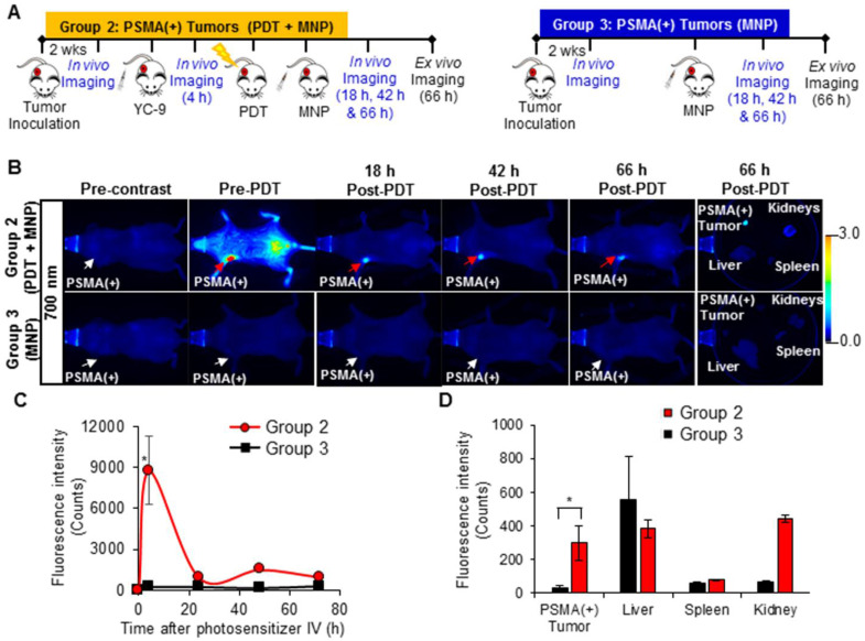
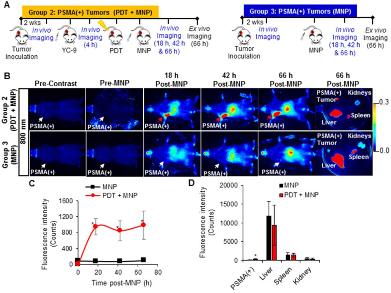
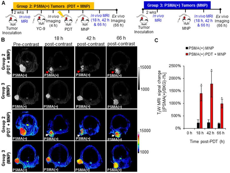
Enhanced vascular permeability in tumors plays an essential role in nanoparticle delivery. Prostate-specific membrane antigen (PSMA) is overexpressed on the epithelium of aggressive prostate cancers (PCs). Here, we evaluated the feasibility of increasing the delivery of PSMA-targeted magnetic nanoparticles (MNPs) to tumors by enhancing vascular permeability in PSMA(+) PC tumors with PSMA-targeted photodynamic therapy (PDT). PSMA(+) PC3 PIP tumor-bearing mice were given a low-molecular-weight PSMA-targeted photosensitizer and treated with fluorescence image-guided PDT, 4 h after. The mice were then given a PSMA-targeted MNP immediately after PDT and monitored with fluorescence imaging and T-weighted magnetic resonance imaging (T-W MRI) 18 h, 42 h, and 66 h after MNP administration. Untreated PSMA(+) PC3 PIP tumor-bearing mice were used as negative controls. An 8-fold increase in the delivery of the PSMA-targeted MNPs was detected using T-W MRI in the pretreated tumors 42 h after PDT, compared to untreated tumors. Additionally, T-W MRIs revealed enhanced peripheral intra-tumoral delivery of the PSMA-targeted MNPs. That finding is in keeping with two-photon microscopy, which revealed higher vascular densities at the tumor periphery. These results suggest that PSMA-targeted PDT enhances the delivery of PSMA-targeted MNPs to PSMA(+) tumors by enhancing the vascular permeability of the tumors.
肿瘤中增强的血管通透性在纳米颗粒递送上起着至关重要的作用。前列腺特异性膜抗原(PSMA)在侵袭性前列腺癌(PC)的上皮细胞中过度表达。在这里,我们通过 PSMA 靶向光动力疗法(PDT)增强 PSMA(+) PC 肿瘤的血管通透性,评估了将 PSMA 靶向磁性纳米颗粒(MNPs)递送到肿瘤中的可行性。PSMA(+) PC3 PIP 荷瘤小鼠给予低分子量 PSMA 靶向光敏剂,并在 4 小时后进行荧光图像引导 PDT 治疗。然后,在 PDT 后立即给予 PSMA 靶向 MNP,并在给药后 18 小时、42 小时和 66 小时进行荧光成像和 T 加权磁共振成像(T-W MRI)监测。未处理的 PSMA(+) PC3 PIP 荷瘤小鼠作为阴性对照。与未处理的肿瘤相比,在 PDT 后 42 小时,T-W MRI 检测到 PSMA 靶向 MNPs 的递送增加了 8 倍。此外,T-W MRI 显示 PSMA 靶向 MNPs 在肿瘤周边的瘤内递增强化。这一发现与双光子显微镜一致,后者显示肿瘤周边的血管密度更高。这些结果表明,PSMA 靶向 PDT 通过增强肿瘤的血管通透性来增强 PSMA 靶向 MNPs 向 PSMA(+)肿瘤的递药。