Rufai Sohaib R, Gore Sri, Handley Sian E, Marmoy Oliver R, Ong Juling, Dunaway David J, Jeelani Noor Ul Owase
Clinical and Academic Department of Ophthalmology, Great Ormond Street Hospital for Children, London, UK.
UCL Great Ormond Street Institute of Child Health, London, UK & Craniofacial Unit, Great Ormond Street Hospital for Children, London, UK.
J Surg Case Rep. 2021 Feb 4;2021(2):rjaa606. doi: 10.1093/jscr/rjaa606. eCollection 2021 Feb.
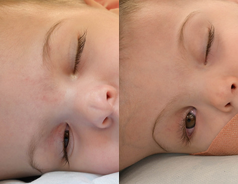
Craniopagus conjoined twins are extraordinarily rare and present unique challenges to the multidisciplinary team. There is a paucity of literature on optimizing neuro-ophthalmologic evaluation in craniopagus twins. Herein, we present our enhanced neuro-ophthalmologic evaluation and management in 17-month-old male craniopagus twins, uniquely using handheld optical coherence tomography (OCT) plus portable slit-lamp biomicroscopy, indirect ophthalmoscopy and modified forced-choice preferential looking assessment. Staged surgical separation was supported by enhanced neuro-ophthalmologic evaluation, detailed radiology, three-dimensional printing and virtual reality simulation. This represents the fourth separation of craniopagus twins by our unit.
颅联体双胞胎极其罕见,给多学科团队带来了独特的挑战。关于优化颅联体双胞胎神经眼科评估的文献很少。在此,我们介绍了对一对17个月大的男性颅联体双胞胎进行的强化神经眼科评估与管理,特别采用了手持光学相干断层扫描(OCT)以及便携式裂隙灯生物显微镜检查、间接检眼镜检查和改良的强迫选择优先注视评估。强化的神经眼科评估、详细的放射学检查、三维打印和虚拟现实模拟为分期手术分离提供了支持。这是我们科室进行的第四例颅联体双胞胎分离手术。