Department of Pathomorphology, Wroclaw Medical University, 50-368 Wroclaw, Poland.
Department of Cell Pathology, Faculty of Biotechnology, University of Wroclaw, 50-383 Wroclaw, Poland.
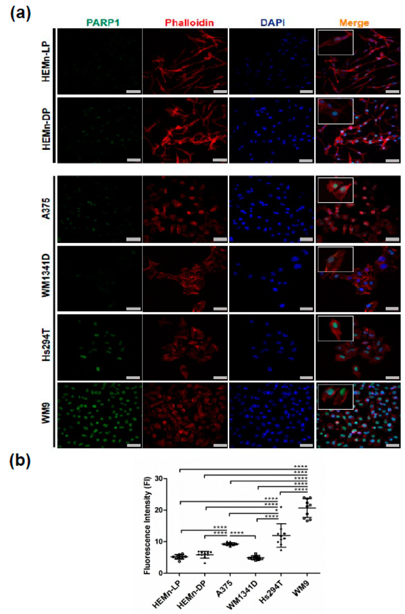
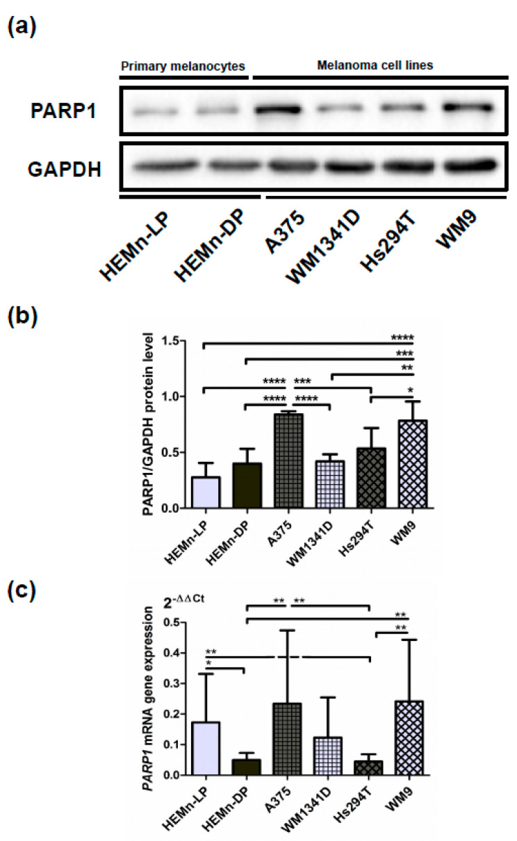
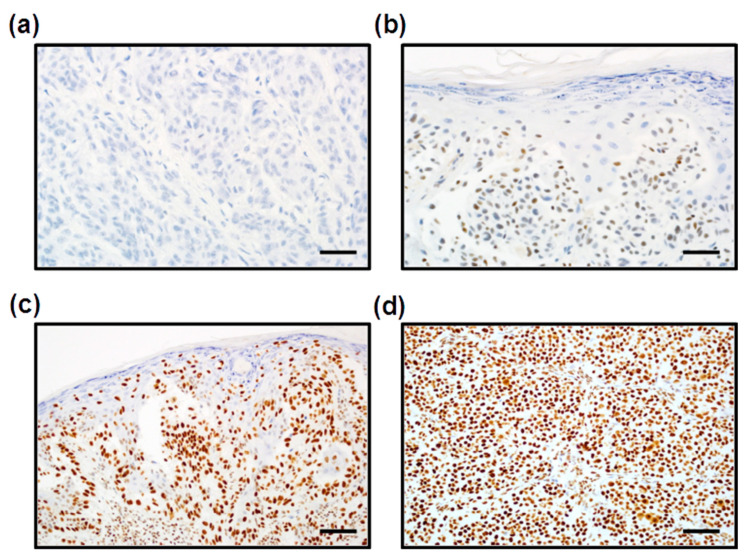
Cells. 2021 Jan 31;10(2):286. doi: 10.3390/cells10020286.
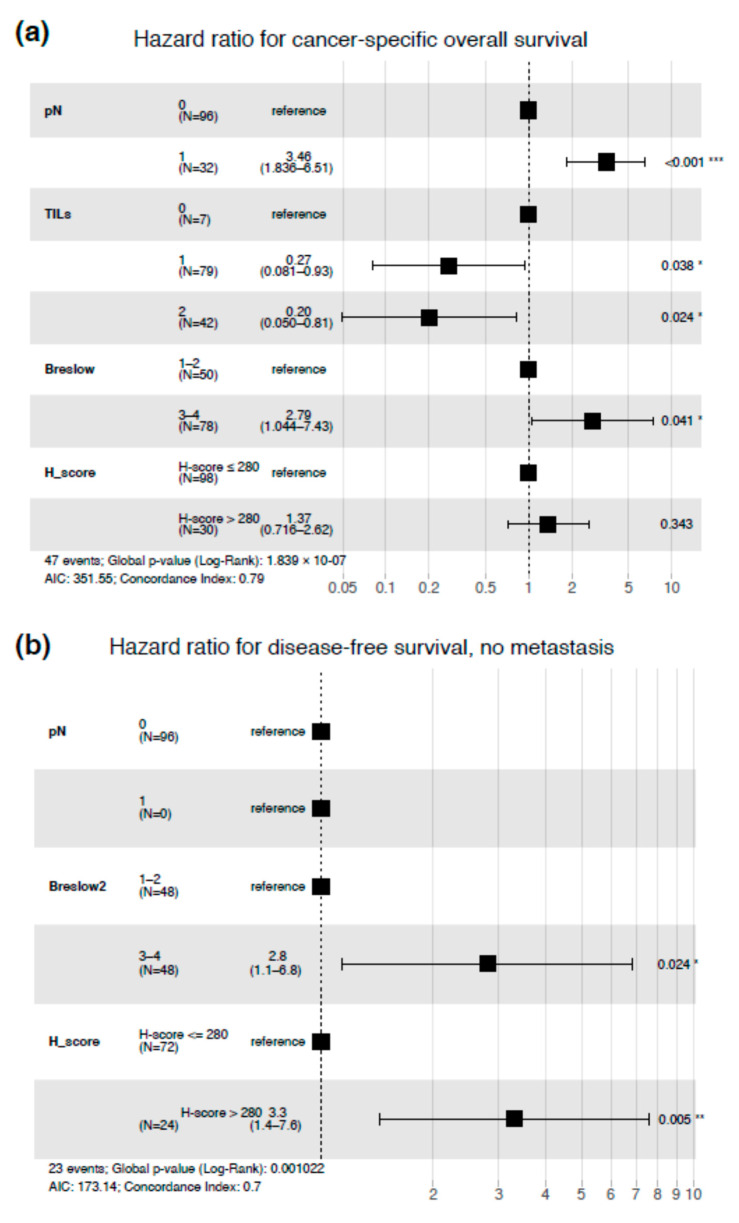
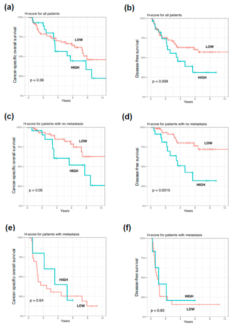
(1) Background: Poly(ADP-ribose) polymerase 1) (PARP1) is a pleiotropic enzyme involved in several cellular processes, e.g., DNA damage repair, regulation of mitosis, and immune response. Little is known about the role of PARP1 in melanoma development and progression. We aimed to investigate the prognostic significance of PARP1 expression in cutaneous melanoma through evaluation of mRNA and protein levels of PARP1 in normal melanocytes and melanoma cell lines, as well as in patients' tissue material from surgical resections. (2) Methods: An in vitro model was based on two types of normal human melanocytes (HEMn-DP and HEMn-LP) and four melanoma cell lines (A375, WM1341D, Hs294T, and WM9). mRNA gene expression was estimated using real-time polymerase chain reaction (RT-PCR), whereas the protein level of PARP1 was evaluated by fluorescence confocal microscopy and then confirmed by Western Blotting analysis. The expression of PARP1 was also assessed by immunohistochemistry in formalin-fixed paraffin-embedded tissues of 128 primary cutaneous melanoma patients and correlated with follow-up and clinicopathologic features. (3) Results: The in vitro study showed that melanoma cells exhibited significantly higher PARP1 expression at mRNA and protein levels than normal melanocytes. High PARP1 expression was also associated with the invasiveness of tumor cells. Elevated nuclear PARP1 expression in patients without nodal metastases strongly correlated with significantly shorter disease-free survival ( = 0.0015) and revealed a trend with shorter cancer-specific overall survival ( = 0.05). High PARP1 immunoreactivity in the lymph node-negative group of patients was significantly associated with higher Breslow tumor thickness, presence of ulceration, and a higher mitotic index ( = 0.0016, = 0.023, and < 0.001, respectively). In patients with nodal metastases, high PARP1 expression significantly correlated with the presence of microsatellitosis ( = 0.034), but we did not confirm the prognostic significance of PARP1 expression in these patients. In the entire analyzed group of patients (with and without nodal metastases at the time of diagnosis), PARP1 expression was associated with a high mitotic index ( = 0.001) and the presence of ulceration ( = 0.036). Moreover, in patients with elevated PARP1 expression, melanoma was more frequently located in the skin of the head and neck region ( = 0.015). In multivariate analysis, high PARP1 expression was an independent unfavorable prognosticator in lymph node-negative cutaneous melanoma patients. (4) Conclusions: In vitro molecular biology approaches demonstrated enhanced PARP1 expression in cutaneous melanoma. These results were confirmed by the immunohistochemical study with clinical parameter analysis, which showed that a high level of PARP1 correlated with unfavorable clinical outcome. These observations raise the potential role of PARP1 inhibitor-based therapy in cutaneous melanoma.
(1) 背景:多聚(ADP-核糖)聚合酶 1(PARP1)是一种参与多种细胞过程的多效酶,例如 DNA 损伤修复、有丝分裂调控和免疫反应。关于 PARP1 在黑色素瘤发展和进展中的作用知之甚少。我们旨在通过评估正常黑素细胞和黑色素瘤细胞系以及手术切除患者组织材料中 PARP1 的 mRNA 和蛋白水平,来研究 PARP1 表达在皮肤黑色素瘤中的预后意义。(2) 方法:体外模型基于两种类型的正常人黑素细胞(HEMn-DP 和 HEMn-LP)和四种黑色素瘤细胞系(A375、WM1341D、Hs294T 和 WM9)。使用实时聚合酶链反应(RT-PCR)估计 mRNA 基因表达,而 PARP1 的蛋白水平则通过荧光共聚焦显微镜评估,然后通过 Western Blotting 分析确认。还通过免疫组织化学法评估了 128 例原发性皮肤黑色素瘤患者福尔马林固定石蜡包埋组织中 PARP1 的表达,并与随访和临床病理特征相关联。(3) 结果:体外研究表明,黑色素瘤细胞在 mRNA 和蛋白水平上的 PARP1 表达明显高于正常黑素细胞。高 PARP1 表达也与肿瘤细胞的侵袭性有关。无淋巴结转移的患者中核 PARP1 表达升高与无病生存期显著缩短(=0.0015)强烈相关,并显示出与癌症特异性总生存期缩短的趋势(=0.05)。在无淋巴结转移的患者中,高 PARP1 免疫反应性与较高的 Breslow 肿瘤厚度、溃疡存在和较高的有丝分裂指数显著相关(=0.0016、=0.023 和 <0.001,分别)。在有淋巴结转移的患者中,高 PARP1 表达与微卫星不稳定性的存在显著相关(=0.034),但我们并未证实这些患者中 PARP1 表达的预后意义。在整个分析组患者(诊断时存在或不存在淋巴结转移)中,PARP1 表达与高有丝分裂指数(=0.001)和溃疡存在相关(=0.036)。此外,在 PARP1 表达升高的患者中,黑色素瘤更常位于头颈部皮肤(=0.015)。在多变量分析中,高 PARP1 表达是无淋巴结转移皮肤黑色素瘤患者的独立不良预后因素。(4) 结论:体外分子生物学方法表明皮肤黑色素瘤中 PARP1 表达增强。这些结果通过临床参数分析的免疫组织化学研究得到了证实,结果表明高水平的 PARP1 与不良的临床结局相关。这些观察结果提示 PARP1 抑制剂为基础的治疗在皮肤黑色素瘤中的潜在作用。