Instituto de Investigaciones Biomédicas Madrid CSIC-UAM. C/Arturo Duperier 4, 28029 Madrid, Spain.
CIBER Enfermedades Cardiovasculares (CIBERCV), Madrid, Spain.
Int J Mol Sci. 2021 Jan 29;22(3):1336. doi: 10.3390/ijms22031336.
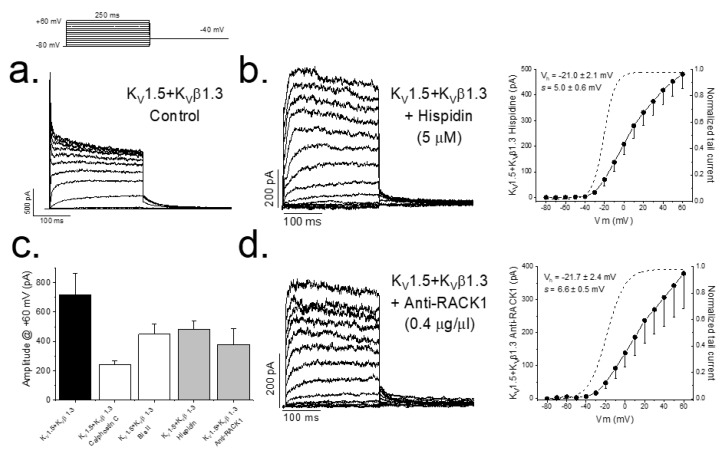
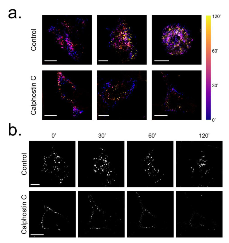
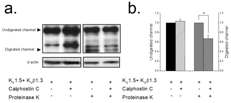
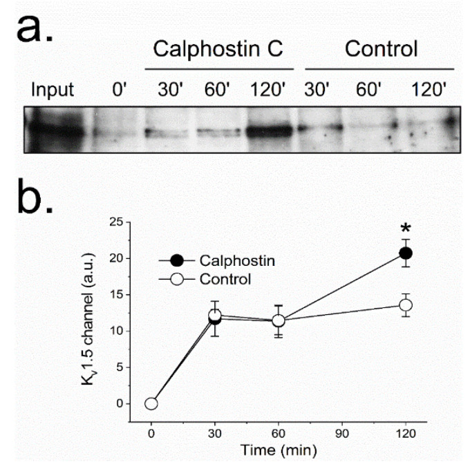
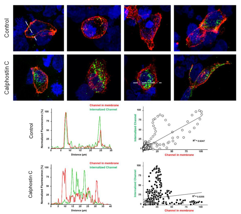
K1.5 channel function is modified by different regulatory subunits. Kβ1.3 subunits assemble with K1.5 channels and induce a fast and incomplete inactivation. Inhibition of PKC abolishes the Kβ1.3-induced fast inactivation, decreases the amplitude of the current K1.5-Kβ1.3 and modifies their pharmacology likely due to changes in the traffic of K1.5-Kβ1.3 channels in a PKC-dependent manner. In order to analyze this hypothesis, HEK293 cells were transfected with K1.5-Kβ1.3 channels, and currents were recorded by whole-cell configuration of the patch-clamp technique. The presence of K1.5 in the membrane was analyzed by biotinylation techniques, live cell imaging and confocal microscopy approaches. PKC inhibition resulted in a decrease of 33 ± 7% of channels in the cell surface due to reduced recycling to the plasma membrane, as was confirmed by confocal microscopy. Live cell imaging indicated that PKC inhibition almost abolished the recycling of the K1.5-Kβ1.3 channels, generating an accumulation of channels into the cytoplasm. All these results suggest that the trafficking regulation of K1.5-Kβ1.3 channels is dependent on phosphorylation by PKC and, therefore, they could represent a clinically relevant issue, mainly in those diseases that exhibit modifications in PKC activity.
K1.5 通道功能可通过不同的调节亚基进行修饰。Kβ1.3 亚基与 K1.5 通道组装,并诱导快速且不完全的失活。PKC 的抑制作用可消除 Kβ1.3 诱导的快速失活,减小 K1.5-Kβ1.3 电流的幅度,并改变其药理学特性,这可能是由于 K1.5-Kβ1.3 通道的运输以 PKC 依赖性方式发生了变化。为了分析该假说,用 K1.5-Kβ1.3 通道转染 HEK293 细胞,并通过膜片钳全细胞配置记录电流。通过生物素化技术、活细胞成像和共聚焦显微镜方法分析膜上 K1.5 的存在。PKC 抑制导致细胞表面的通道减少 33±7%,这是由于向质膜的循环减少所致,这一点通过共聚焦显微镜得到了证实。活细胞成像表明,PKC 抑制几乎消除了 K1.5-Kβ1.3 通道的循环,导致通道在细胞质中积累。所有这些结果表明,K1.5-Kβ1.3 通道的运输调节依赖于 PKC 的磷酸化,因此,它们可能是一个具有临床意义的问题,特别是在那些 PKC 活性发生改变的疾病中。