Department of Dermatology and Venereology, National Clinical Research Center for Skin and Immune Diseases, Research Center for Medical Mycology, Beijing Key Laboratory of Molecular Diagnosis on Dermatoses, Peking University First Hospital, Beijing, People's Republic of China.
Emerg Microbes Infect. 2021 Dec;10(1):331-341. doi: 10.1080/22221751.2021.1889931.
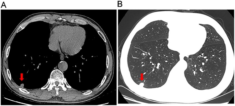
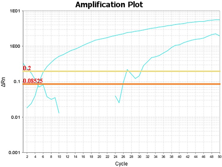
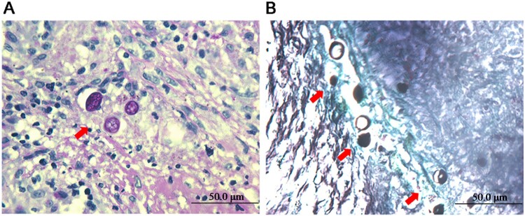
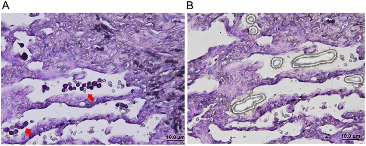
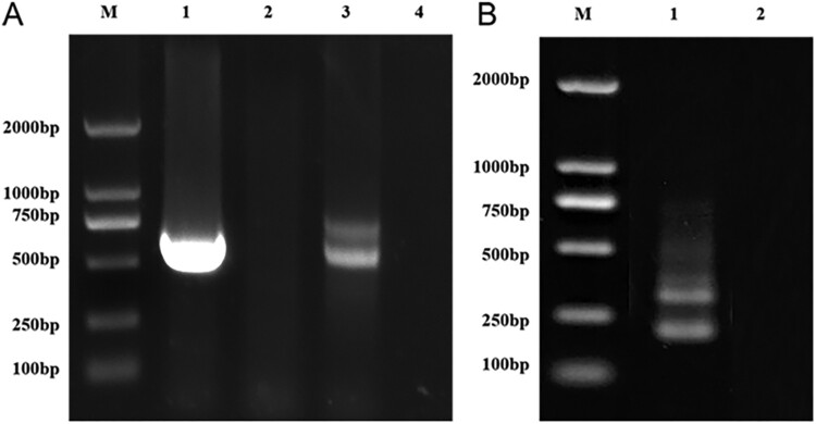
Coccidioidomycosis is endemic to California, Arizona, and Mexico. In recent years, the reported cases of coccidioidomycosis have increased in nonendemic regions. Here, we reported a case of imported pulmonary coccidioidomycosis in a Chinese patient. A 63-year-old man presented with dry cough and fatigue for 6 months, and a computed tomography scan revealed a solitary nodule in the right lower lung and small nodules in both lungs. The diagnosis of coccidioidomycosis was initially confirmed by histopathologic examination. The pathogen spp. was identified by laser capture microdissection (LCM) combined with subsequent molecular techniques based on the positive histopathologic features. Additionally, we reviewed 47 reported cases of coccidioidomycosis in China. The number of reported cases is increasing, and the incidence of disseminated infection has exhibited a trend of shifting towards healthy young adults in China. Since clinical presentations and imaging findings lack specificity, a majority of domestic cases of coccidioidomycosis were initially misdiagnosed as tumours or tuberculosis. Moreover, the diagnosis of endemic mycoses may be challenging because of their rarity and the limited availability of diagnostic tests. The diagnosis was mainly confirmed by histopathological examination. The species involved were identified based on positive cultures in only 4 cases. To our knowledge, this is the first study to use LCM and molecular techniques to identify spp. in the histopathologically positive but uncultivable specimen. Comparing with previous reported studies, LCM combined with nucleic acid amplification techniques improve the ability of species identification for the timely diagnosis of coccidioidomycosis.
球孢子菌病流行于加利福尼亚、亚利桑那和墨西哥。近年来,非流行地区报告的球孢子菌病病例有所增加。在此,我们报告了一例中国患者的输入性肺球孢子菌病。一名 63 岁男性因干咳和乏力就诊 6 个月,计算机断层扫描显示右下肺单发结节和双肺小结节。球孢子菌病的诊断最初通过组织病理学检查确认。通过激光捕获显微切割(LCM)联合后续基于阳性组织病理学特征的分子技术,鉴定出病原体 种。此外,我们复习了中国 47 例球孢子菌病病例。报告病例数呈上升趋势,中国播散性感染的发病率向健康年轻成人转移。由于临床表现和影像学表现缺乏特异性,国内大多数球孢子菌病病例最初被误诊为肿瘤或结核病。此外,由于其罕见性和诊断检测的局限性,地方性真菌病的诊断可能具有挑战性。诊断主要通过组织病理学检查确认。仅 4 例通过阳性培养鉴定出涉及的种。据我们所知,这是首例使用 LCM 和分子技术鉴定组织学阳性但无法培养标本中 种的研究。与以往报道的研究相比,LCM 结合核酸扩增技术提高了种鉴定的能力,有助于及时诊断球孢子菌病。